Yanlış küvet malzemesi seçimi, spektroskopik verileri fark edilmeden bozar — ve çoğu araştırmacı, bu hatayı ancak sonuçlar açıklanamaz hale geldikten sonra fark eder.
UV-Vis spektroskopisinde küvet malzemesi önemsiz bir konu değildir; bu, absorbans değerlerinin numunenin kimyasal özelliklerini mi yoksa cihaz kaynaklı sapmaları mı yansıttığını doğrudan belirleyen temel bir değişkendir. Bu makale, kuvarsın UV-Vis ölçümleri için referans malzeme olmasını sağlayan optik, yapısal, kimyasal ve işlevsel özelliklerini incelemekte ve aynı zamanda tüm spektral aralıkta cam, plastik ve safir alternatifleriyle performansını sistematik olarak karşılaştırmaktadır.
Spektroskopide malzeme seçimi, cihazın numuneyi barındıran kapta ne tür özellikler aradığını anlamakla başlar. Bir UV-Vis spektrofotometre 190 nm ile 800 nm arasındaki dalga boylarını tararken, ışık yolundaki her optik bileşen — küvet dahil — radyasyonu emmeden, saçmadan veya floresan göstermeden geçirmelidir. Bu aralıktaki herhangi bir dalga boyunda ışın demetini bozan bir küvet, yalnızca yazılımla yapılan sonradan işlemeyle düzeltilemeyen sistematik hataya yol açar.
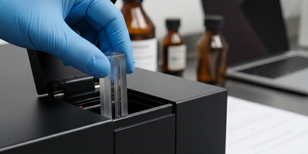
Optik Şeffaflık Penceresi, Küvet Malzemesinin Seçimini Belirler
Her optik malzeme radyasyonu seçici bir şekilde geçirir ve bu seçiciliğin sınırları, bir küvetin UV-Vis çalışmaları için bilimsel açıdan uygun olup olmadığını ya da bu çalışmalarla temelde uyumsuz olup olmadığını belirler.
Optik Malzemelerde İletim Penceresi Nedir?
Bir geçirgenlik aralığı, bir malzemenin elektromanyetik ışınımı önemli bir zayıflama olmaksızın geçmesine izin verdiği dalga boyu aralığını ifade eder. Zayıflama, birbiriyle rekabet halindeki üç olgudan kaynaklanır: emilim, yansıma ve saçılma, bunların her biri dalga boyuna bağlı bir şekilde sinyal kaybına katkıda bulunur.
Atom düzeyinde, emilim, gelen foton enerjisinin, malzemenin moleküler veya kristal yapısı içindeki elektronik temel durumlar ile uyarılmış durumlar arasındaki enerji aralığına denk geldiğinde gerçekleşir. Geniş elektronik bant aralığına sahip malzemeler — yani elektronları uyarılmış durumlara yükseltmek için gereken enerjinin UV fotonlarının enerjisini aştığı malzemeler — geniş, kısa dalga boylu geçirgenlik aralıkları sergiler. Buna karşılık, geçiş metali iyonları, konjuge organik yapılar veya yapısal kusurlar içeren malzemeler, UV radyasyonunu kolayca emen ara elektronik enerji durumlarına sahiptir ve karakteristik kesme dalga boylarının altındaki geçirgenliği etkili bir şekilde engeller.
Küvet seçimine ilişkin pratik sonuç doğrudan ortaya çıkmaktadır: Absorpsiyon eşiği ölçümün spektral aralığı içinde kalan herhangi bir malzeme, kendi absorpsiyon profilini numune sinyaline ekleyecek ve bu da doğru bir taban çizgisi düzeltmesini imkânsız hale getirecektir.
UV-Vis Spektral Aralığı ve Zorlu Optik Gereksinimleri
UV-Vis spektral aralığı geleneksel olarak 190 nm ile 800 nm arası, derin UV (190–280 nm), yakın UV (280–400 nm) ve görünür ışık (400–800 nm) bölgelerine ayrılır. Her bir alt bölge, küvet malzemesinin şeffaflığı konusunda kendine özgü gereksinimler ortaya koyar.
Görünür bölge (400–800 nm) nispeten esnektir; hem cam hem de kuvars bu aralıkta yeterli düzeyde ışık geçirir; bu da tamamen görünür ışıkta gerçekleştirilen kolorimetrik analizler için malzeme seçimini daha az kritik hale getirir. Bununla birlikte, yakın UV bölgesi (280–400 nm), cam ve çoğu polimerin sınırlarını ortaya çıkarmaya başlar; çünkü bu malzemelerin emilim sınırları 320 nm'nin altından yukarı doğru uzanır. Derin UV bölgesi (190–280 nm) ise en zorlu bölgedir: Ticari açıdan kullanışlı üçten az küvet malzemesi — erimiş silika kuvars, UV sınıfı sentetik kuvars ve safir — 220 nm'nin altında yeterli şeffaflığı korur.
280 nm’de protein miktar tayini, 260 nm’de nükleik asit miktar tayini, 215 nm’de peptit bağı absorpsiyonu ve 250–290 nm aralığında aromatik amino asit karakterizasyonu, tümü yakın UV bölgesinde veya bu bölgenin yakınında yer almaktadır. Rutin laboratuvar spektroskopisinin önemli bir bölümünü oluşturan bu uygulamalar için, Küvet malzemesinin 320 nm altındaki dalga boylarında şeffaflığı mutlak bir gerekliliktir.
UV-Vis Ölçümleri, Kuvars Küvetin Optik Özelliklerinden Nasıl Yararlanır?
Sadece optik şeffaflık, UV-Vis ölçümlerinin neden bu kadar büyük ölçüde fayda sağladığını tam olarak açıklamaz kuvars küvet yapısı. Bu avantaj, moleküler yapıdan üretim hassasiyetine ve yüzey kalitesine kadar uzanmaktadır.
Füzyon Silikanın Moleküler Yapısı ve UV Şeffaflığı
UV-Vis kuvars küvetlerinde kullanılan başlıca malzeme olan erimiş silika, köşelerini paylaşan [SiO₄] tetrahedrlerinden oluşan sürekli ve rastgele bir ağ halinde düzenlenmiş, tamamen silikon dioksitten (SiO₂) oluşan amorf bir katıdır. Uzun menzilli düzenli bir kafes yapısına sahip olan kristal kuvarsın aksine, fused silika, periyodik yapısal düzenlilikten yoksundur, bu da çift kırılmayı ortadan kaldırır ve malzemeyi tüm yönlerde optik olarak izotropik hale getirir.
Si-O bağının elektronik yapısı, erimiş silikanın UV şeffaflığının temelini oluşturur. Yüksek saflıkta erimiş silikanın bant aralığı yaklaşık olarak 8,9 eV, bu da vakum UV bölgesinde 140 nm civarında bir emilim başlangıcına karşılık gelir. 190 nm'deki fotonlar yaklaşık 6,5 eV enerji taşır — bu değer, Si-O bant aralığı boyunca elektronları uyarmak için gereken eşiğin oldukça altındadır — yani laboratuvar spektroskopisiyle ilgili dalga boylarındaki UV radyasyonu, saf erimiş silikadan elektronik emilim olmaksızın geçer. Bu durum, katkı maddesi oksitlerinin 350 nm'ye kadar uzun dalga boylarındaki UV radyasyonunu emen bant aralığı altı elektronik durumlar yarattığı çok bileşenli camlarla keskin bir tezat oluşturmaktadır.
SiCl₄’ün alev hidrolizi veya plazma CVD yöntemi ile üretilen sentetik erimiş silika, 1 ppm’nin altında hidroksil (OH) konsantrasyonları ve 20 ppb’nin altında metalik safsızlık seviyeleri sağlar, her ikisi de UV şeffaflığının korunması açısından hayati öneme sahiptir. Doğal kuvars kristalleri, arıtma işleminden sonra bile, 245 nm civarında (oksijen eksikliği merkezleriyle ilişkili) ve 214 nm civarında (E' merkezleriyle ilişkili) emilim bantlarına yol açan eser miktarda safsızlıklar barındırır; bu da onları derin UV uygulamaları için sentetik erimiş silikadan daha düşük performanslı hale getirir.
Bir Kuvars Küvetin Derin UV’den Yakın Kızılötesiye Kadar Olan Geçiş Aralığı
Yüksek saflıkta erimiş silika, yaklaşık 170 nm ile 2.700 nm arası, başka hiçbir uygun maliyetli optik malzemenin tam olarak eşleştiremediği bir spektral aralığı kapsar. Özellikle UV-Vis aralığında (190–800 nm), 10 mm yol uzunluğuna sahip UV sınıfı erimiş silika küvetin geçirgenlik değerleri genellikle 200 nm'de 85% ve 250 nm'de 92%, bu kayıpların esas olarak kütle emilimi yerine iki hava-cam arayüzündeki Fresnel yansımasından kaynaklandığı belirtilmelidir.
Ticari sınıfta üç farklı tipte erimiş silika küvet, esas olarak hidroksil içeriği ve buna bağlı IR emilimi açısından birbirinden ayrılır. UV sınıfı erimiş silika (düşük OH, < 10 ppm OH) 250 nm’nin altında optimum geçirgenlik sağlar; bu da onu derin UV spektroskopisi için uygun bir seçim haline getirir. Standart sınıf erimiş silika (yüksek OH, 400–1.000 ppm OH), 245 nm civarındaki OH absorpsiyon üst tonları nedeniyle UV geçirgenliğinde hafif bir azalma gösterir, ancak 220 nm üzerindeki çoğu yakın UV uygulaması için kabul edilebilir bir performans sergiler. IR sınıfı erimiş silika, bazı UV performansından ödün vererek 2.000–3.500 nm aralığında geçirgenliği optimize eder.
Biyoloji laboratuvarlarında en yaygın iki UV-Vis uygulaması olan nükleik asit ve protein miktar tayini için, 10 mm yol uzunluğuna sahip UV sınıfı erimiş silika küvetler, 260 nm ve 280 nm dalga boylarında 0,01 AU’nun altında baz hattı absorbans değerleri sağlar, geniş bir konsantrasyon aralığında doğru niceleme için gerekli ölçüm aralığını sağlar.
Kuvars Küvet Sınıfları Arasında Kırılma İndisi Tekdüzeliği ve Yüzey Kalitesi
589 nm'de (sodyum D çizgisi) erimiş silikanın kırılma indisi yaklaşık olarak 1.458, 193 nm’de 1,534’ten 1.064 nm’de 1,440’a kadar öngörülebilir bir şekilde değişen bir dağılım profiline sahiptir. Mutlak kırılma indisi değerinden daha önemli olan, küvetin optik penceresi boyunca bu değerin uzamsal homojenliğidir: Yüksek kaliteli erimiş silika ham parçaları, ±5 × 10⁻⁶’dan daha iyi bir kırılma indisi homojenliği sergilerler, bu sayede küvet duvarlarının neden olduğu dalga cephesi bozulmasının, cihazın fotometrik hassasiyetine kıyasla ihmal edilebilir düzeyde olmasını sağlar.
Yüzey kalitesi, küvet duvarlarındaki saçılma kayıplarını doğrudan belirler. Erimiş silikanın optik sınıfta parlatılmasıyla, yüzey pürüzlülüğü değerleri şu seviyenin altına düşer: 0,5 nm RMS (kare ortalaması), bu da UV-Vis aralığı boyunca yüzey saçılma kayıplarını 0,11 TP3T’nin altında tutar. Bu spesifikasyona göre parlatma işlemi, giderek daha ince aşındırıcıların kullanıldığı çok aşamalı alıştırma işleminin ardından ziftle parlatma veya manyetoreolojik son işlem gerektirir — bu işlemler optik imalata özgüdür ve standart laboratuvar cam malzemesi üretiminden farklıdır.
Çift taraflı cilalı küvetler — ışık demetine dik olan sadece iki yüzün optik olarak işlendiği — bu tür yüzeyler, standart absorbans ölçümleri için yeterlidir. Dört kenarlı cila küvetleri, burada dört dikey yüzeyin tamamı aynı optik kaplamaya sahip olup, bunlar floresan spektroskopisi için gereklidir, dairesel dikroizm (CD)1, ve ışınların yan yüzlerden içeri girdiği veya dışarı çıktığı durumlarda yapılan optik dönme ölçümleri.
Kuvars Küvetin Optik Performans Özeti
| Parametre | UV Sınıfı Erimiş Silika | Standart Erimiş Silika | IR Sınıfı Erimiş Silika |
|---|---|---|---|
| İletim Aralığı (nm) | 170–2.700 | 190–2.700 | 220–3.500 |
| OH İçeriği (ppm) | < 10 | 400–1.000 | < 10 |
| 200 nm'de geçirgenlik (%) | > 85 | 75–85 | 60–75 |
| 260 nm'de geçirgenlik (%) | > 92 | > 90 | > 88 |
| 589 nm'de Kırılma İndeksi | 1.458 | 1.458 | 1.458 |
| Yüzey Pürüzlülüğü RMS (nm) | < 0.5 | < 0.5 | < 0.5 |
| Metalik Safsızlıklar (ppb) | < 20 | < 50 | < 20 |
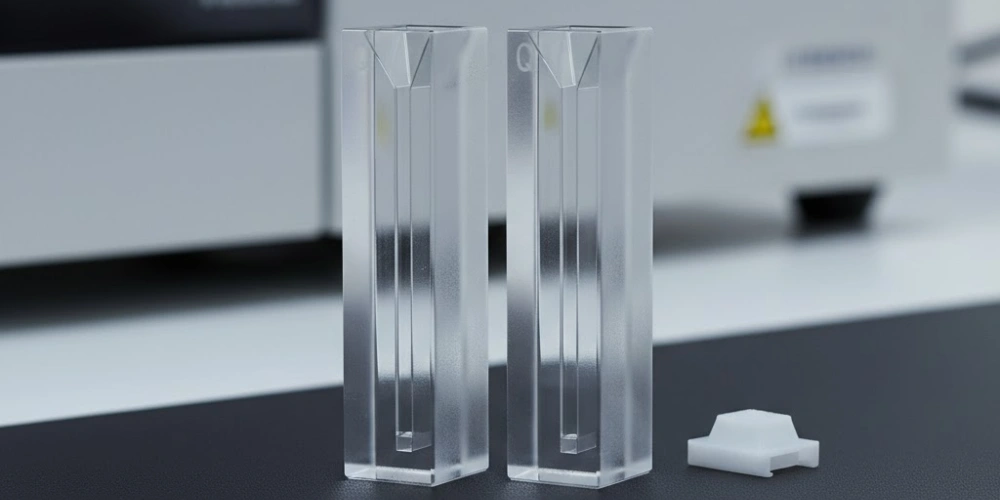
UV-Vis Kuvars Küvetlerinde Işın Yolu Uzunluğunun Seçimi ve Bunun Ölçüm Doğruluğuna Etkisi
Malzeme şeffaflığının ötesinde, bir UV-Vis kuvars küvetinin ışık yol uzunluğu, absorbans ölçümlerinin hem doğruluğunu hem de doğrusal dinamik aralığını etkileyen en önemli geometrik parametredir.
Beer-Lambert Yasası ve Yol Uzunluğuna Olan Doğrusal Bağımlılık
Beer-Lambert yasası, absorbans, numune konsantrasyonu ve optik yol uzunluğu arasındaki temel ilişkiyi ifade eder: A = ε × c × l; burada A absorbans (boyutsuz), ε molar zayıflama katsayısı (L mol⁻¹ cm⁻¹), c molar konsantrasyon (mol L⁻¹) ve l yol uzunluğu (cm) değerlerini temsil eder. Bu yasa, sabit yol uzunluğunda absorbans ile konsantrasyon arasında ve sabit konsantrasyonda absorbans ile yol uzunluğu arasında kesin bir doğrusal ilişki öngörür; ancak bu doğrusallık yalnızca belirli bir aralıkta geçerlidir.
Absorbans değerleri yaklaşık olarak şu değeri aştığında, Beer-Lambert doğrusallığından sapmalar belirgin hale gelir 1,5 AU (3,2%’nin altındaki geçirgenliğe karşılık gelir). Yüksek absorbans değerlerinde, spektrofotometre içindeki parazit ışık — numuneden geçmeden dedektöre ulaşan radyasyon — algılanan sinyalin orantılı olarak daha büyük bir kısmını oluşturur ve bu da görünür absorbansın gerçek değerinin altında bir düzlükte kalmasına neden olur. Ayrıca, yüksek çözünen madde konsantrasyonlarında, emici türler arasındaki moleküller arası etkileşimler, etkin molar zayıflama katsayısını değiştirerek doğrusal olmaktan kimyasal sapmalara yol açar. Her iki etki de gerçek konsantrasyonu sistematik olarak düşük göstermekte olup, 2,0 AU’nun üzerindeki absorbans değerlerinde hatalar 5–151 TP3T’ye ulaşmaktadır..
Yol uzunluğunu azaltmak, yüksek konsantrasyonlu numunelerde doğrusallığı yeniden sağlamak için fiziksel açıdan kesin bir yöntemdir. Yol uzunluğunu 10 mm’den 5 mm’ye yarıya indirmek, sabit konsantrasyonda ölçülen absorbansı yarıya indirir ve numune seyreltmesine gerek kalmadan ölçümleri doğrusal aralığa geri getirir — bu, numune hacimleri sınırlı olduğunda veya seyreltme işleminin çözelti dengelerini bozacağı durumlarda kritik bir avantajdır.
Kuvars Küvetlerdeki Standart Işın Yolları ve Bunlara Ait Uygulamalar
Üreticiler, yaklaşık olarak şu yol uzunluğu aralığını kapsayan UV-Vis kuvars küvetleri üretmektedir: üç büyüklük mertebesi, 0,1 mm'den 100 mm'ye kadar, analitik uygulamalarda karşılaşılan tüm numune konsantrasyonlarını kapsayacak şekilde.
Kısa yol uzunluğuna sahip küvetler — 0,1 mm, 0,2 mm ve 0,5 mm — seyreltilmemiş protein stokları, konsantre boya çözeltileri ve proses konsantrasyonlarındaki farmasötik formülasyonlar gibi yüksek konsantrasyonlu numuneler için kullanılır. 0,1 mm ışık yolu uzunluğunda, 10.000 L mol⁻¹ cm⁻¹ molar zayıflama katsayısına sahip bir numune, yaklaşık Tipik bir küresel protein için 150 mg/mL — standart 10 mm küvetlerle ulaşılamayan bir aralık. Bu 10 mm ışık yolu uzunluğu Küvet, rutin UV-Vis ölçümleri için evrensel bir standarttır ve çoğu laboratuvar numunesi için yaklaşık 0,05–1,5 AU’luk pratik bir çalışma absorbans aralığı sunar. Uzun yol uzunluğuna sahip küvetler 20 mm, 50 mm ve 100 mm hassasiyeti, aromatik kirleticiler açısından analiz edilen çevresel su numuneleri, ultra-seyreltilmiş farmasötik safsızlık standartları ve ekolojik araştırmalardaki düşük konsantrasyonlu kromoforik türler dahil olmak üzere, eser konsantrasyonlu numunelere genişletmek.
Sınırlı Hacimli UV-Vis Analizi için Mikro ve Alt-Mikro Kuvars Küvetleri
Özellikle genomik, proteomik ve tek hücre biyolojisinde yaygın olarak görülen numune hacmi kısıtlamaları, tam optik performansı korurken önemli ölçüde daha küçük hacimler gerektiren mikro ve sub-mikro kuvars küvet formatlarının geliştirilmesine yol açmıştır.
Standart bir 10 mm yol uzunluğuna sahip kuvars küveti için yaklaşık olarak 3,0–3,5 mL ışın yolunun üzerindeki optik hazneyi doldurmak için gerekli numune miktarı. Yarı mikro küvetler bu gereksinimi 1,4–1,7 mL 10 mm’lik yol uzunluğunu koruyarak iç hazne genişliğini daraltarak. Mikro küvetler, hacim gereksinimlerini daha da azaltarak 0,6–0,7 mL, ve mikro altı formatlar ise sadece 70 µL, spektrofotometrenin optik geometrisine uyumlu, Z boyutu (küvet tabanının üzerindeki ışın merkezinin yüksekliği) hassas bir şekilde belirlenmiş, son derece dar bir iç hazne (genellikle 3 mm × 3 mm kesitli) tasarlanarak elde edilmiştir.
Z boyutu, hayati önem taşıyan bir uyumluluk parametresidir: Çoğu masaüstü UV-Vis spektrofotometresinde ışın merkezi 8,5 mm küvet tabanının üstünde bulunurken, bazı cihazlarda 15 mm veya 20 mm’lik Z boyutları kullanılır. Küvetin Z boyutu ile cihazın ışın yüksekliği arasındaki uyumsuzluk, ışının numune hacminin bir kısmını kaçırmasına neden olur ve bu da, bağımsız bir doğrulama yapılmadıkça gerçek düşük konsantrasyon sinyallerinden ayırt edilemeyen, anormal derecede düşük absorbans okumalarına yol açar.
Kuvars Küvet Yolu Uzunluğu ve Hacim Referansı
| Yol uzunluğu (mm) | Tipik Numune Hacmi (mL) | Birincil Uygulama |
|---|---|---|
| 0.1 | 0.05-0.15 | Yüksek konsantrasyonlu protein stokları, API çözeltileri |
| 0.5 | 0,10–0,30 | Konsantre biyolojik özler |
| 1 | 0,40–0,70 | Orta konsantrasyonlu numuneler |
| 10 | 3.00–3.50 | Genel rutin UV-Vis ölçümleri |
| 20 | 6.00–7.00 | Çevre numunelerini seyreltin |
| 50 | 15,0–17,0 | İz miktarda kirletici maddelerin analizi |
| 100 | 30,0–35,0 | Ultra-eser konsantrasyon ölçümleri |
UV-Vis Deney Düzenekleri için Kuvars Küvetlerdeki Geometrik Tasarım Çeşitliliği
Yol uzunluğu ve hacim özellikleri, birincil optik eksen boyunca neler olduğunu ele alır; ancak küvet gövdesinin geometrisinin kendisi, belirli deney düzeneklerinde ölçüm kalitesini etkileyen ek değişkenler ortaya çıkarır.
Dört Yüzlü Parlak Küvetler ile İki Yüzlü Parlak Küvetlerin Karşılaştırılması
Çift taraflı cilalı küvetler, yalnızca uyarma ışınına dik olan iki yüzeyde — yani ölçüm ışınının fiilen geçtiği yüzeylerde — optik olarak düz ve çiziksiz yüzeyler sunar. Kalan iki yan yüzey ise mat bir yüzeye sahip olacak şekilde taşlanmıştır; bu yüzeyler mekanik stabilite ve kullanım açısından yeterlidir, ancak ışın geçişi açısından optik olarak uygun değildir. Çift taraflı cila küvetleri, standart absorbans ölçümleri için tamamen yeterlidir burada ışın, cilalı bir yüzeyden girip, tek bir doğrusal optik eksen boyunca karşıdaki cilalı yüzeyden çıkar.
Dört kenarlı cilalı küvetler, dört dikey yüzünün tamamında aynı optik yüzey kalitesine sahiptir; bu sayede ışınım, saçılma kaybı olmaksızın herhangi bir yüzeyden girip çıkabilir. Bu yapı, aşağıdakiler için vazgeçilmezdir: floresan spektroskopisi, burada uyarma ışınımı bir yüzeyden girer ve yayılan floresan, bitişik bir yüzeyden 90° açıyla toplanır. Bu durum aynı şekilde şunlar için de gereklidir: dairesel dikroizm (CD) spektroskopisi, burada etkileşim geometrisi, optik olarak homojen yüzeyler aracılığıyla gerçekleştirilen hassas polarizasyon kontrolüne bağlıdır ve optik rotasyon ölçümleri için kullanılır. İki yüz yerine dört yüzün parlatılmasından kaynaklanan ek üretim maliyeti — genellikle bir 30–50% premium eşdeğer çift taraflı cilalama özelliklerine kıyasla — yalnızca ölçüm geometrisi yanal optik erişim gerektirdiğinde haklıdır.
Pratik bir tanımlama yöntemi: İki taraflı cilalı küvetler, eğik ışık altında incelendiğinde genellikle yan duvarlarında gözle görülür bir opaklık veya buzlu bir görünüm sergilerken, dört taraflı cilalı küvetler ise her açıdan eşit derecede şeffaf görünür.
Siyah Duvarlı Kuvars Küvetler ve Floresan Ölçümlerinde Parazit Işığın Engellenmesi
Standart dört kenarlı cila küvetleri, her yönden optik olarak erişilebilir olmalarına rağmen, floresan ölçümlerinde belirli bir yapay etki yaratır: iç küvet duvarlarından yansıyan uyarma ışınımı, emisyon dedektörüne ulaşır, gerçek floresan spektrumunun üzerine bir arka plan sinyali bindirerek. Bu artefakt, özellikle uyarma dalga boyuna yakın emisyon dalga boylarında — suyun Raman saçılma zirvesi bölgesinde ve floresan emisyon bandının başlangıç kısmında — oldukça belirgindir.
Siyah cidarlı kuvars küvetler, şu yöntemi uygulayarak bu yapay etkiyi ortadan kaldırır: iki yan yüzeye ve arka yüzeye opak siyah bir kaplama, sadece ön yüzeyi (uyarma girişi) ve 90°'lik emisyon yüzeyini şeffaf ve cilalı bırakarak. Siyah kaplama, yansıyan ve saçılan uyarma ışınımını emisyon dedektörüne ulaşmadan önce emerek, parazit ışık arka planını 10–100 katlık faktörler floresan deneylerinde standart dört kenarlı cilalı küvetlere kıyasla. Siyah cidarlı bir küvetin kuvars gövdesi, UV sınıfı erimiş silikadan oluşmaya devam eder; yalnızca dış yüzey kaplaması farklıdır.
Floresan spektroskopisinde siyah cidarlı küvet yerine standart şeffaf cidarlı küvet kullanmanın pratik sonucu, yüksek ve spektral olarak yapılandırılmış bir arka plan sinyalidir. Bu durum, hassasiyeti azaltır, uyarma dalga boyunun yaklaşık 30–50 nm aralığındaki dalga boylarında emisyon spektrumlarını bozar ve zayıf floresan gösteren numunelerde kantitatif doğruluğu olumsuz etkiler.
Kuvars Küvet Tasarım Çeşitleri ve Uygulamaları
| Tasarım Tipi | Cilalı Yüzler | Dağınık Işık Kontrolü | Birincil Uygulama |
|---|---|---|---|
| Çift taraflı cila | 2 (ön ve arka) | Standart | UV-Vis absorbansı |
| Dört taraflı cilalama | 4 (hepsi dikey) | Standart | Floresans, CD spektroskopisi |
| Siyah duvarlı | 2 (ön + emisyon yüzü) | Geliştirilmiş | Düşük arka planlı floresan |
| Akışlı | 2 veya 4 | Standart | HPLC dedektörleri, sürekli akış |
| Silindirik | Sürekli eğri | Sınırlı | Özel dairesel dikroizm |
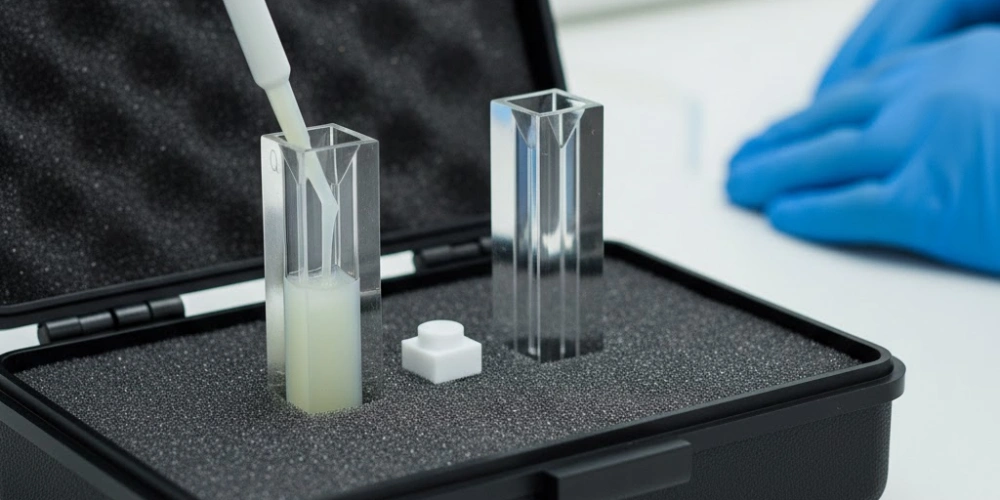
Farklı Numune Matrislerinde UV-Vis Kuvars Küvetlerinin Kimyasal Direnç Profilleri
Füzyon silika kuvarsın spektroskopik üstünlüğüne, kimyasal dayanıklılık açısından da aynı derecede önemli bir avantaj eşlik etmektedir — bu avantaj, bir küvetin optik yüzeylerinde bozulma meydana gelmeden güvenle barındırabileceği numune yelpazesini belirlemektedir.
Eritilmiş Silikanın Asit ve Organik Çözücülerle Uyumluluğu
Füzyon silikanın asitlere ve organik çözücülere karşı kimyasal direnci, doğrudan Si-O bağ ağının termodinamik kararlılığından kaynaklanmaktadır. Yaklaşık 452 kJ/mol değerindeki Si-O bağı ayrışma enerjisi Bu değer, çok bileşenli camlardaki çoğu metal-oksijen bağının değerini aşmaktadır; bu da, erimiş silikanın, geleneksel laboratuvar cam malzemelerine kolayca zarar veren reaktiflere neden dayanıklı olduğunu açıklamaktadır.
Güçlü mineral asitler — tüm konsantrasyonlardaki hidroklorik asit, yaklaşık 70% konsantrasyonuna kadar sülfürik asit, nitrik asit ve oda sıcaklığındaki fosforik asit dahil — tipik laboratuvar maruz kalma süreleri boyunca erimiş silika yüzeylerine ölçülebilir bir şekilde zarar vermez. Biyokimyada rutin olarak kullanılan sulu asidik tamponlar (pH 3–6 aralığındaki sitrat, asetat, fosfat ve MES tamponları) da benzer şekilde zararsızdır. Organik çözücüler de aynı derecede geniş bir uyumluluk aralığına sahiptir: etanol, metanol, izopropanol, aseton, asetonitril, dimetil sülfoksit (DMSO), diklorometan, kloroform, toluen ve tetrahidrofuran (THF) hepsi, şişme, sızıntı veya optik yüzey bozulmasına yol açmadan erimiş silika kuvetlerle uyumludur.
Önemli bir pratik ayrım Füzyon silika kuvetleri, polimerik kuvet malzemelerinden ayırır: PMMA veya polistiren kuvetlerin anında çatlamasına, bulanıklaşmasına veya çözünmesine neden olan organik çözücüler — özellikle klorlu çözücüler, ketonlar ve aromatik hidrokarbonlar — kuvars tarafından sorunsuz bir şekilde işlenir; bu da erimiş silika kuvars küvetleri, susuz çözücü sistemlerinde UV-Vis ölçümleri için tek pratik seçenek haline getirir.
Hidroflorik Asit ve Konsantre Alkalilerin Önemli İstisnası
Geniş kimyasal uyumluluğuna rağmen, erimiş silika kuvarsın, laboratuvar uygulamalarında istisnasız olarak dikkate alınması gereken iki açıkça tanımlanmış kimyasal zayıflığı bulunmaktadır.
Hidroflorik asit (HF), SiO₂ ile şu stokiyometrik reaksiyon yoluyla reaksiyona girer: SiO₂ + 4HF → SiF₄↑ + 2H₂O, uçucu maddeler üreterek silikon tetraflorür2 ve su. Bu reaksiyon, 0,1% gibi düşük HF konsantrasyonlarında ve oda sıcaklığında bile ölçülebilir hızlarda ilerler ve temasın başlamasından itibaren birkaç dakika içinde kuvars küvetlerin cilalı optik yüzeylerini aşındırır. Yüzey aşınması, temizlikle giderilemeyen kalıcı saçılma kayıplarına ve dalga cephesi bozulmasına yol açar; HF'ye maruz kalan bir kuvet, onarılamaz şekilde hasar görmüş olarak değerlendirilmeli ve kullanımdan kaldırılmalıdır. Konsantre sodyum hidroksit (NaOH > 30%) ve potasyum hidroksit (KOH > 20%), erimiş silikayı daha yavaş bir çözünme mekanizmasıyla aşındırır — hidroksit iyonları, Si-O-Si bağlarını hidrolitik olarak koparır; çözünme hızları yaklaşık olarak saatte 0,1–1 µm oda sıcaklığında konsantre alkali içinde. 100 °C’nin üzerindeki sıcaklıklarda konsantre fosforik asit de erimiş silikayı, sıcaklıkla birlikte keskin bir şekilde artan oranlarda aşındırır.
HF, konsantre alkali veya sıcak konsantre fosforik asit gerektiren numuneler için uygun alternatif kaplar, politetrafluoroetilen (PTFE) veya perfloroalkoksi (PFA) kaplardır., bunların hiçbiri bu reaktiflerle reaksiyona girmiyor.
Erimiş Silika Kuvetlerinin Kimyasal Uyumluluğu
| Reaktif Sınıfı | Örnek | Uyumluluk | Notlar |
|---|---|---|---|
| Seyreltilmiş mineral asitler | HCl, H₂SO₄, HNO₃ | ✓ Uyumlu | Tüm konsantrasyonlar, ortam sıcaklığı |
| Organik çözücüler | EtOH, Aseton, DMSO, CHCl₃ | ✓ Uyumlu | Şişkinlik veya optik bozulma görülmez |
| Sulu tamponlar (pH 3–9) | PBS, HEPES, sitrat | ✓ Uyumlu | Standart biyolojik pH aralığı |
| Seyreltilmiş alkali çözeltiler (< 5%) | NaOH, KOH | ⚠ Dikkat | Yavaş pozlama; pozlama süresini en aza indirin |
| Konsantre alkali maddeler | NaOH > 30% | ✗ Uyumsuz | Birkaç saat içinde yüzeyden çözünme |
| Hidroflorik asit | HF (herhangi bir konsantrasyon) | ✗ Uyumsuz | Anında geri dönüşümsüz aşındırma |
| Sıcak konsantre H₃PO₄ | > 85%, > 100 °C | ✗ Uyumsuz | SiO₂ saldırısının termal hızlanması |
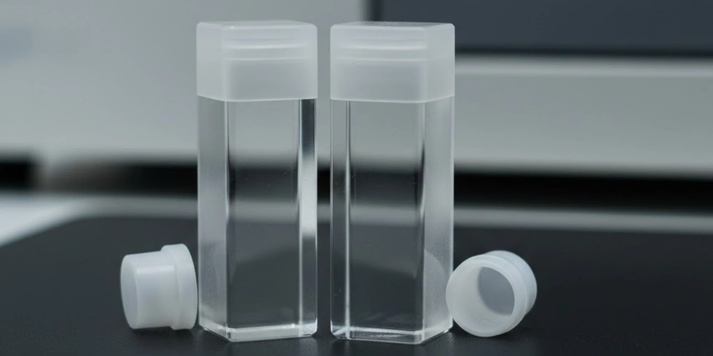
320 nm Altındaki UV-Vis Ölçümlerinde Cam Küvetlerin Neden Yetersiz Kaldığı
Cam küvetler, eğitim laboratuvarlarında ve görünür ışık aralığındaki kolorimetride hâlâ yaygın olarak kullanılmaktadır; ancak ultraviyole bölgesindeki spektroskopik sınırlamaları, bu küvetleri analitik uygulamaların büyük bir kısmı için bilimsel açıdan uygun olmayan hale getirecek kadar ciddi boyuttadır.
Borosilikat ve Soda-Kireç Camının Kimyasal Bileşimi
Ticari laboratuvar cam küvetleri, borosilikat cam veya soda-kireç camından üretilir — bu iki çok bileşenli silikat bileşimi, önemli oranda silika dışı ağ oluşturucular ve modifiye ediciler içerir.
Borosilikat cam (Schott DURAN ve Corning Pyrex gibi örneklerde görüldüğü üzere) yaklaşık olarak 81% SiO₂, 13% B₂O₃, 4% Na₂O ve 2% Al₂O₃ ağırlıkça. Termal genleşme katsayısını azaltmak ve termal şok direncini artırmak amacıyla bor trioksit (B₂O₃) eklenir; ancak bor-oksijen yapısal birimleri, saf SiO₂’de bulunmayan UV aralığında elektronik geçişlere yol açar. Soda-kireç bardağı genellikle şunları içerir 72% SiO₂, 14% Na₂O, 9% CaO ve 5% MgO — optik performanstan ziyade işlenebilirlik ve maliyet açısından optimize edilmiş bir bileşim. Ağ düzenleyici oksitler (Na₂O, CaO, MgO), Si-O ağını bozarak, UV ışınlarını emen kusur merkezleri oluşturan köprü oluşturmayan oksijen bölgeleri yaratır.
Her iki cam türünde de eser miktarda metal kirlilikleri — özellikle standart optik camda 50–200 ppm konsantrasyonlarda bulunan Fe²⁺ ve Fe³⁺ iyonları — yoğun UV emilim bantları oluşturur. Fe³⁺, 225 nm ve 320 nm civarında emilim bantları üretir; bu, ligand alanı d-d geçişleri3 ve yük transferi geçişlerine katkıda bulunurken, Fe²⁺ ise 200 nm civarında emilime katkıda bulunur. 10 mm'lik bir yol uzunluğu boyunca 100 ppm'nin altındaki konsantrasyonlarda bile, bu demir absorpsiyon bantları 280 nm'de 0,1–0,5 AU'luk absorbans katkıları üretir — bu değerler, seyreltilmiş biyolojik numunelerin sinyallerini bastıracak büyüklüktedir.
Camın UV Emme Kesme Noktası ve Spektroskopik Sonuçları
Bir küvet malzemesinin ultraviyole emilim kesme noktası, geleneksel olarak, malzemenin kendine özgü absorbansının aşağıdakine eşit olduğu dalga boyu olarak tanımlanır: 1,0 AU 10 mm’lik bir yol uzunluğu için — bu noktada, gelen radyasyonun yalnızca 10%’si iletilir ve sinyal-gürültü oranı, nicel ölçümün güvenilirliğini ortadan kaldıracak bir düzeye düşer.
Borosilikat cam için bu kesme dalga boyu şu aralıkta yer alır: 290 ve 320 nm belirli cam bileşimine ve demir içeriğine bağlı olarak. Soda-kireç camı için bu sınır değeri genellikle 320–350 nm. Bu eşik değerleri, borosilikat cam küvetlerin 280 nm’de (aromatik absorbans yoluyla protein miktar tayini), 260 nm’de (nükleik asit miktar tayini), 254 nm (UV sterilizasyon izleme) ve 214–220 nm (peptit bağı absorbansı ve düşük dalga boylu protein miktar tayini) dalga boylarındaki ölçümler için kullanılamaz. Bir cam küvet 280 nm'lik bir protein tayini için kullanıldığında, camın kendisi yaklaşık 0,3–0,8 AU nominal olarak aynı teknik özelliklere sahip tek tek küvetler arasında farklılık gösteren — tek bir boş ölçümle düzeltilemeyen, partiye bağlı sistematik bir hata.
Spektroskopik sonuçlar birbirini takip eder: sıcaklığa bağlı cam emicilik kaymalarının yol açtığı taban çizgisi kayması, camın emilim sınırlarına karşılık gelen dalga boylarında görülen görünür emilim tepe noktaları (bunlar numunenin kromoforlarına yanlış bir şekilde atfedilebilir) ve tüm niceleme işlemlerini Beer-Lambert eğrisinin üstteki, doğrusal olmayan kısmına zorlayan daralmış doğrusal dinamik aralık.
UV Dalga Boylarında Camda Oluşan Otofloresans ve Saçılma Artifaktları
Doğrudan emilimin ötesinde, cam küvetler UV aralığında ikincil optik bozulmalara yol açarak ölçüm doğruluğunu daha da olumsuz etkiler.
Camın otofloresansı — UV uyarımı sonrasında görünen ışınımın kendiliğinden yayılması — cam üretimi sırasında camın içine karışan yapısal kusur merkezleri ve organik kirleticiler içindeki elektronik geçişlerden kaynaklanır. Borosilikat cam 280 nm dalga boyunda ışınlandığında, zirvesi yaklaşık 400–450 nm, cam partileri arasında değişen kuantum verimlerine sahiptir. Standart bir tek ışınlı UV-Vis spektrofotometrede, bu floresan, monokromatörün geçiş bandının emisyon spektrumuyla çakıştığı dalga boylarında tespit edilen sinyale katkıda bulunur ve numune absorbansında görünür bir azalmaya neden olur — bu, uyarma yoğunluğuyla doğrusal olmayan bir şekilde orantılı olan ve kuvars küvette sadece çözücü kullanılarak yapılan boş ölçümlerde görülmeyen bir artefakt.
Camdaki mikroskobik kalıntılar — hapsolmuş gaz kabarcıkları, karışmamış eriyik bölgeler ve kristalit çökeltileri — şu işlevi görür: Mie saçılma merkezleri UV radyasyonu için. Ölçüm dalga boyuyla karşılaştırılabilir çaplara sahip küresel parçacıklardan kaynaklanan Mie saçılımı (UV radyasyonu için 100–300 nm), daha kısa dalga boylarına doğru dik bir şekilde artan ve kolloidal parçacıkların absorpsiyon profilini taklit eden, dalga boyuna bağlı bir arka plan oluşturur. Uygulamada, 220 nm’de yapılan ölçümler için kullanılan bir cam küvet, saçılmadan kaynaklanan ve 0,5 AU — birçok seyreltilmiş biyolojik numunede, gerçek numune absorbansından daha yüksektir.
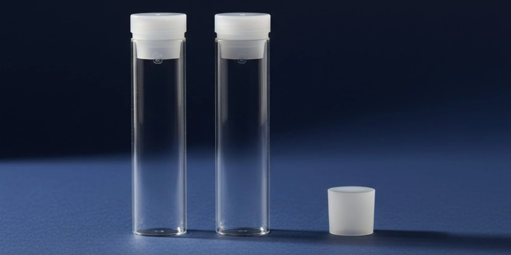
Kuvars Cam, Plastik ve Safir Küvetler Arasında İletim Performansı Karşılaştırması
Uygun küvet malzemesinin seçilmesi, laboratuvar spektroskopistlerinin kullanabileceği dört malzeme sınıfının tümü — erimiş silika kuvars, borosilikat cam, polimetilmetakrilat (PMMA) ve safir — arasında, UV-Vis ölçümlerinin özel gereklilikleri ışığında yapılan sistematik bir karşılaştırma gerektirir.
Erimiş silika kuvars Bu malzeme, dört malzeme arasında en geniş pratik geçirgenlik aralığını, en yüksek kimyasal uyumluluğu ve en iyi optik yüzey kararlılığını sunar; bunun karşılığında birim üretim maliyeti de daha yüksektir. 170 nm ile 2.700 nm arasındaki geçirgenliği ve 260 nm'de < 0,01 AU temel absorbansı, onu diğer tüm malzemelerin karşılaştırma ölçütü olarak kabul edilen referans malzeme haline getirir.
Borosilikat cam 320 nm'nin üzerinde ve tüm görünür ışık aralığında (400–800 nm) kuvarsla karşılaştırılabilir bir geçirgenlik sağlar; bu da onu kolorimetrik analizler, görünür dalga boylarında izlenen enzim kinetiği ve 320 nm'nin altındaki UV ışınlarına erişim gerektirmeyen her türlü ölçüm için uygun ve maliyet etkin hale getirir. 290–320 nm civarındaki UV kesme noktası ve UV ışınlaması altında otofloresansa yatkınlığı, onu yakın UV bölgesi için uygun olmayan bir malzeme haline getirir.
PMMA ve polistiren plastik küvetler, UV kesme dalga boyları PMMA için 300–320 nm ve Polistiren için 340–360 nm — bu cihazları görünür ışık aralığındaki kolorimetriyle sınırlayan kısıtlamalar. Başlıca avantajları fiyat ve kullanım kolaylığıdır: Tek kullanımlık protokollerin zorunlu olduğu klinik ve yüksek verimli ortamlarda çapraz kontaminasyon endişelerini ortadan kaldırırlar. Organik çözücüler plastik küvetleri anında çözer veya çatlatır ve optik sınıf olmayan yüzeyleri önemli ölçüde saçılma gösterir. Safir (Al₂O₃) küvetler yaklaşık olarak 145 nm ile 5.500 nm arası Olağanüstü kimyasal direnç ve mekanik sertliğe (Mohs 9) sahip olmaları nedeniyle, 160 nm altındaki vakum UV uygulamalarında erimiş silikadan teknik açıdan daha üstündürler. Bununla birlikte, safirin trigonal kristal yapısından kaynaklanan çift kırılma özelliği, polarimetrik ölçümleri zorlaştırır ve imalatındaki zorluklar, kullanımını özel araştırma uygulamalarıyla sınırlar.
Küvet Malzemesi Geçirgenliği ve Uyumluluk Karşılaştırması
| Mülkiyet | Erimiş Silika Kuvars | Borosilikat Cam | PMMA Plastik | Safir |
|---|---|---|---|---|
| İletim Aralığı (nm) | 170–2.700 | 320–2.500 | 300–900 | 145–5.500 |
| 1,0 AU / 10 mm'de UV kesme değeri (nm) | < 175 | 290–320 | 300–320 | < 150 |
| 260 nm'nin altında kullanılabilir | ✓ Evet | ✗ Hayır | ✗ Hayır | ✓ Evet |
| Organik Solvent Direnci | Mükemmel | İyi | Zayıf | Mükemmel |
| HF Direnci | ✗ Hayır | ✗ Hayır | ✓ Evet | ✓ Evet |
| UV ışığı altında otofloresans | İhmal edilebilir | Önemli | Orta düzeyde | İhmal edilebilir |
| Yüzey Sertliği (Mohs) | 7 | 6-7 | 3 | 9 |
| Yeniden kullanılabilir | ✓ Evet | ✓ Evet | ✗ Hayır | ✓ Evet |
| Birefringence | Hiçbiri | Hiçbiri | Hiçbiri | Mevcut |
Küvet Malzemesi veya Durumundan Kaynaklanan Yaygın Ölçüm Hataları
Doğru küvet malzemesi seçilmiş olsa bile, küvetin durumundan kaynaklanan ölçüm hataları — çizikler, kalıntı kirlilik, yanlış yönlendirme ve hapsolmuş kabarcıklar — UV-Vis spektroskopisinde veri kalitesi sorunlarının sürekli bir kaynağıdır.
-
Temel çizgi kayması ve sıfırdan farklı boş numune absorbansı küvetle ilgili en sık karşılaşılan hatalardır. Yüzeyinde kir birikmiş bir kuvars küvet boş ölçüm için kullanıldığında, kaydedilen baz çizgisi kirin absorbansını sıfır olarak dahil eder; bu da sonraki tüm numune ölçümlerinde gerçek absorbansın aynı miktarda düşük gösterilmesine neden olur. Kuvars küvetin optik yüzeyinde bulunan bir protein tabakası, 280 nm'de 0,05–0,2 AU görünür absorbans — standart bir Bradford veya doğrudan UV analizinde protein konsantrasyonunun 10–50% oranında yanlış hesaplanmasına yol açacak kadar büyük bir hata. Buna karşılık, sıcaklığa bağlı bazal kayma, küvetten ziyade numuneden kaynaklanır: sulu çözeltilerin kırılma indisi yaklaşık olarak °C başına −0,0001, küvet arayüzlerindeki Fresnel yansıma kayıplarını değiştirerek ve sıcaklık stabilize olduğunda tersine çevrilebilir olmasıyla ayırt edilen yavaş bir absorbans kayması oluşturur.
-
Anormal derecede yüksek absorbans değerleri Bilinen numune konsantrasyonlarıyla uyuşmayan değerler, genellikle numunenin artan absorpsiyonundan ziyade, küvetin optik yüzeyinde çiziklerin neden olduğu saçılmadan kaynaklanır. Işın kesitini kesen tek bir çizik, görünen absorbansı 0,05–0,5 AU, çizik derinliğine ve genişliğine bağlı olarak; daha kısa dalga boylarında saçılma katkısı keskin bir şekilde artar. Çizik kaynaklı saçılmayı gerçek numune emiliminden ayırt etmek için, aynı boş çözelti kullanılarak görünüşte anormal sonuç veren küveti temiz bir referans küvetiyle karşılaştırmak gerekir; çizik kaynaklı saçılma, kalıcı bir taban çizgisi kayması olarak kalırken, gerçek numune emilimi numune konsantrasyonuna göre değişir.
-
Ölçüm tekrarlanabilirliğinin yetersizliği — Aynı numunelerin tekrarlı ölçümlerinde 1–2%’yi aşan varyasyon katsayısı — genellikle küvetin takılma yönündeki tutarsızlıklardan kaynaklanır. Çoğu küvet tam olarak kare şeklinde değildir: duvar kalınlığındaki farklılıklar ±0,01-0,05 mm Karşılıklı yüzler arasındaki mesafe, ışına hangi yüzün yöneltildiğine bağlı olarak etkin yol uzunluğunu değiştirir. Tutarlı bir yerleştirme yönünün belirlenmesi (laboratuvar kalemi ile işaretlenerek veya üreticinin yönlendirme işaretiyle hizalanarak), genellikle yönelim kaynaklı absorbans değişkenliğini 0,3%’nin altına indirir.
-
Kabarcık bozulmaları normalde düzgün davranış gösteren dalga boylarında ani ve büyük absorbans artışlarına — genellikle 1,0 AU’yu aşan — neden olur. Işın kesitinin sadece bir kısmını kaplayan bir kabarcık bile, gelen radyasyonun neredeyse tamamını dedektörden uzaklaştırarak, numunenin neredeyse tam emilimini simüle eder. Kabarcıklar, numunelerin soğuk depodan oda sıcaklığına getirilmesi sırasında çözeltiden çıkan çözünmüş gazdan, dar çaplı pipetler aracılığıyla yapılan türbülanslı numune aktarımından ve yeterince kurutulmamış küvetlerde hapsolmuş durulama çözücüsü kalıntılarından kaynaklanır. Ölçümden önce oda sıcaklığına nazikçe ısıtma, numunenin ışın demetine doğrudan değil, küvetin duvarı boyunca yavaşça verilmesi ve kullanımlar arasında iyice kurutulması kabarcık oluşumunu güvenilir bir şekilde önler.
Her Ölçüm İşlemi Öncesinde UV-Vis Kuvars Küvetinin Bütünlüğünün Doğrulanması
UV-Vis kuvars küvetini kantitatif ölçümlere kullanmadan önce kısa bir doğrulama prosedürü uygulamak, deney veri setleri genelinde düzeltilmemiş sistematik hataların birikmesini önler.
-
Temel bulaşma doğrulaması En bilgilendirici ön ölçüm kontrolüdür. Küveti HPLC sınıfı suyla (veya deneyde kullanılacak saf çözücüyle) doldurup hava referansına karşı 190 nm'den 350 nm'ye kadar tarama yapmak, hem kalıntı kontaminasyonu (karakteristik dalga boylarında yüksek absorbans) hem de yüzey saçılımını (daha kısa dalga boylarına doğru düzgün bir şekilde yükselen yüksek baz çizgisi) ortaya çıkarır. HPLC sınıfı suyla doldurulmuş temiz bir UV sınıfı erimiş silika kuvet, aşağıda belirtilen değerin altında bir absorbans göstermelidir 200 nm'de 0,05 AU, 230 nm'de 0,02 AU'nun altında ve 260 nm'de 0,01 AU'nun altında standart spektrofotometre koşulları altında bir hava boşluğu ile karşılaştırılarak. Bu eşik değerlerin üzerindeki sapmalar, ya kalıntı kirliliğine (ek temizlik gerektirir) ya da optik yüzey hasarına (küvetin değiştirilmesini gerektirir) işaret eder.
-
Eğik ışık altında gözle inceleme spektrofotometrik temel kontrolünü tamamlayarak, belirgin spektral emilim özellikleri oluşturmasa da saçılmaya neden olan çizik desenlerini, bulanıklığı ve mekanik kırıkları ortaya çıkarır. Küveti bir floresan tüpüne veya fiber optik ışık kaynağına yaklaşık 30° açıyla tutarak ve optik yüzeyleri geçirgen ışık altında incelediğinizde, çizikler parlak doğrusal çizgiler olarak görülür; bulanıklık cam gövde içinde dağınık bir parıltı olarak görünür; mekanik çentikler ise küvetin köşelerinde veya kenarlarında keskin kenarlı parlak bölgeler olarak ortaya çıkar. İçinde çizikler bulunan herhangi bir küvet optik yüzeyin merkezindeki 80% — spektrofotometre ışınının geçtiği bölge — kantitatif ölçümler için hizmet dışı bırakılmalıdır.
-
Eşleştirilmiş çift doğrulama Çift ışınlı spektrofotometreler, ayrı numune ve referans küvetleriyle kullanıldığında bu işlem gereklidir. Her iki küveti de aynı boş çözeltiyle doldurup, 200–400 nm aralığında birinin diğerine göre absorbansını ölçmek, bunların fotometrik eşdeğerliğini nicel olarak belirler. Eşleşen bir çift, absorbans farklarının Tüm dalga boyu aralığı boyunca 0,005 AU; şu sayıyı aşan çiftler Ölçüm aralığı içindeki herhangi bir dalga boyunda 0,51 TP3T geçirgenlik farkı yeniden eşleştirilmeli veya değiştirilmelidir; zira bu uyumsuzluk, tek bir boş ölçümle sıfırlanamayan, dalga boyuna bağlı bir taban çizgisi hatasına yol açmaktadır.
-
Değiştirme kriterleri UV-Vis kuvars küvetlerinin kullanım ömrü, kronolojik yaş veya kullanım sayısından ziyade optik performansa göre belirlenir. Temel geçiş testi ve görsel incelemeyi geçen bir küvet, ne kadar süredir kullanımda olursa olsun güvenilir ölçümler vermeye devam eder. Buna karşılık, kapsamlı bir temizliğe rağmen temel geçiş testini geçemeyen — belirli bir değerin üzerinde kalıcı olarak yüksek absorbans gösteren — bir küvet HPLC sınıfı çözücüde 260 nm'de 0,05 AU — optik yüzeyinde kalıcı bir bozulma meydana gelmiştir ve nicel UV-Vis çalışmalarında artık kullanılmamalıdır.
Sonuç
UV-Vis küvetlerinde malzeme seçimi, tüm spektral aralık boyunca veri bütünlüğü üzerinde doğrudan etkileri olan bir karardır. Eritilmiş silika kuvars, cam, plastik ve çoğu rakip malzemeden ayrılır; çünkü moleküler yapısı — 8,9 eV'lik bir elektronik bant aralığına sahip sürekli bir SiO₂ ağı — 170 nm'den 2.700 nm'ye kadar emilim, otofloresans veya asitler ve organik çözücüler varlığında yüzey bozulması olmadan 170 nm ile 2.700 nm arasında ışını geçirir. Cam küvetler, geçiş metali safsızlıkları, yapısal kusurlar ve UV emilimi, baz çizgisi kayması ve floresan artefaktlarına neden olan çok bileşenli oksit bileşimleri nedeniyle 320 nm'nin altında yetersiz kalır. Yol uzunluğu, geometri ve temizlik gereksinimlerine uygun doğru küvet seçimi, UV-Vis spektroskopisi için ikincil bir konu değildir — bu, her kantitatif sonucun dayandığı fiziksel temeldir.
SSS
Herhangi bir UV-Vis ölçümü için cam küvet kullanılabilir mi?
Cam küvetler, tamamen 320 nm’nin üzerinde gerçekleştirilen ölçümler için kullanılabilir — görünür aralıktaki kolorimetri, 400–800 nm aralığında izlenen enzim kinetik analizleri ve absorbansa dayalı bulanıklık ölçümleri. Bu küvetler, 280 nm'de protein tayini, 260 nm'de nükleik asit tayini veya yakın UV bölgesinde aromatik veya peptid bağı absorbansına dayalı herhangi bir analiz dahil olmak üzere, 320 nm'nin altındaki dalga boylarına erişim gerektiren hiçbir ölçüm için uygun değildir.
Çoğu UV-Vis uygulaması için standart olarak hangi yol uzunluğuna sahip kuvars küveti kullanılır?
10 mm yol uzunluğuna sahip kuvars küveti, çoğu biyolojik ve kimyasal analizde tipik olarak karşılaşılan numune konsantrasyonları için yaklaşık 0,05–1,5 AU’luk pratik bir absorbans çalışma aralığı sağladığı için evrensel standarttır; tablolarda verilen molar zayıflama katsayıları değerlerinde varsayılan yol uzunluğuna doğrudan karşılık gelir (bu katsayılar geleneksel olarak L mol⁻¹ cm⁻¹ birimlerinde rapor edilir; burada 1 cm = 10 mm) ve neredeyse tüm ticari masaüstü spektrofotometrelerin optik geometrisiyle uyumludur.
Bir kuvars küveti ne sıklıkla değiştirilmelidir?
Değiştirme sıklığı, takvim süresine göre değil, optik performansa göre belirlenir. Temel çizgi geçirgenlik testini geçen — HPLC sınıfı çözücüde 260 nm'de 0,05 AU'dan az değer gösteren — ve merkezi optik yüzey alanında çizik bulunmayan bir kuvars küveti, süresiz olarak kullanımda kalabilir. Bu eşik değerin üzerinde kalıcı olarak yüksek temel çizgi absorbansı, kapsamlı temizlikten sonra da devam ediyorsa, bu durum geri dönüşü olmayan yüzey hasarını teyit eder ve değiştirme gerekliliğini gösterir.
UV-Vis analizlerinde kuvarsın performansını aşan herhangi bir kuvet malzemesi var mı?
Safir (Al₂O₃), erimiş silikadan daha geniş bir geçirgenlik aralığına sahiptir; bu aralık, vakum UV’deki yaklaşık 145 nm’den orta kızılötesi dalga boyundaki 5.500 nm’ye kadar uzanır. Bununla birlikte, 190–800 nm aralığıyla sınırlı laboratuvar UV-Vis uygulamaları için, erimiş silika kuvars, safir ile eşdeğer performans gösterirken, polarimetrik ve dairesel dikroizm ölçümlerini zorlaştıran bir özellik olan safirin doğasında bulunan çift kırılma özelliğini ortadan kaldırır; bu da UV sınıfı erimiş silika kuvarsı, UV-Vis spektroskopik uygulamalarının büyük çoğunluğu için pratik açıdan en uygun seçenek haline getirir.
Referanslar:
-
Dairesel dikroizm spektroskopisi, kiral moleküller tarafından sol ve sağ yönlü dairesel polarize ışığın farklı emilimlerini ölçer; bu işlemde, polarizasyon durumunun bütünlüğünü korumak için optik olarak homojen küvet yüzeyleri gereklidir.↩
-
Silikon tetraflorür, hidroflorik asit ile silikon dioksit arasındaki reaksiyonun sonucu olarak ortaya çıkan uçucu bir gazdır ve bu maddenin oluşumu, erimiş silika yüzeylerinin HF’ye maruz kalması durumunda geri dönüşümsüz bir aşınmaya yol açar.↩
-
Ligand alanı geçişleri, çevreleyen ligandların elektrostatik alanı içinde d-orbital enerji seviyelerinin ayrışmasıyla ortaya çıkan ve geçiş metali iyonları içindeki elektronik uyarımlardır; bu geçişler, metal içeren camlarda karakteristik UV ve görünür ışık emilim bantları oluşturur.↩