Le choix d'un matériau inadapté pour la cuvette fausse les données spectroscopiques sans qu'on s'en aperçoive — et la plupart des chercheurs ne se rendent compte de l'erreur que lorsque les résultats deviennent inexplicables.
Le matériau de la cuvette n'est pas un aspect secondaire en spectroscopie UV-Vis ; il s'agit d'une variable fondamentale qui détermine directement si les mesures d'absorbance reflètent la composition chimique de l'échantillon ou s'il s'agit d'artefacts liés à l'instrument. Cet article examine les propriétés optiques, structurelles, chimiques et opérationnelles qui font du quartz le matériau de référence pour les mesures UV-Vis, tout en comparant systématiquement ses performances à celles d’autres matériaux (verre, plastique et saphir) sur l’ensemble du spectre.
Le choix du matériau en spectroscopie commence par la compréhension des exigences réelles de l’instrument vis-à-vis du récipient contenant l’échantillon. Lorsqu’un spectrophotomètre UV-Vis balaye des longueurs d’onde comprises entre 190 nm et 800 nm, chaque composant optique situé sur le trajet optique — y compris la cuvette — doit transmettre le rayonnement sans l’absorber, le diffuser ni émettre de fluorescence. Une cuvette qui perturbe le faisceau à n'importe quelle longueur d'onde comprise dans cette plage introduit une erreur systématique qui ne peut être corrigée par le seul post-traitement logiciel.
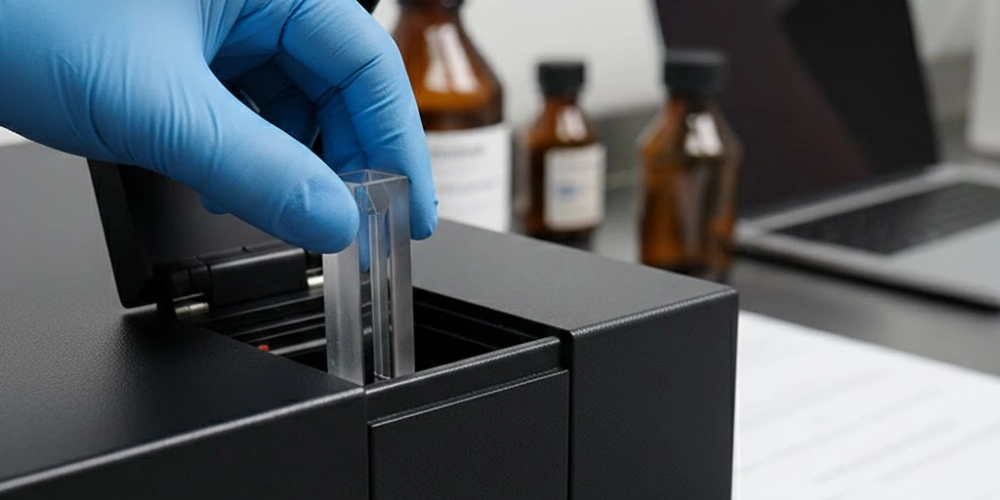
La transparence optique de la fenêtre détermine le choix du matériau de la cuvette
Chaque matériau optique transmet le rayonnement de manière sélective, et ce sont les limites de cette sélectivité qui déterminent si une cuvette est scientifiquement adaptée aux analyses UV-Vis ou si elle est fondamentalement incompatible avec celles-ci.
Qu'est-ce qu'une fenêtre de transmission dans les matériaux optiques ?
Une fenêtre de transmission désigne la gamme de longueurs d'onde dans laquelle un matériau laisse passer le rayonnement électromagnétique sans atténuation significative. L'atténuation résulte de trois phénomènes concurrents : l'absorption, la réflexion et la diffusion, qui contribuent chacune à la perte de signal d'une manière dépendante de la longueur d'onde.
Au niveau atomique, l’absorption se produit lorsque l’énergie du photon incident correspond à la bande interdite entre les états fondamentaux et les états excités des électrons au sein de la structure moléculaire ou cristalline du matériau. Les matériaux présentant une large bande interdite — c’est-à-dire dont l’énergie nécessaire pour exciter les électrons dépasse l’énergie des photons UV — présentent des fenêtres de transmission larges et à courte longueur d’onde. À l’inverse, les matériaux contenant des ions de métaux de transition, des structures organiques conjuguées ou des défauts structurels possèdent des états d’énergie électroniques intermédiaires qui absorbent facilement le rayonnement UV, bloquant ainsi efficacement la transmission en dessous de leurs longueurs d’onde de coupure caractéristiques.
L'implication pratique pour le choix de la cuvette est directe: tout matériau dont le seuil d'absorption se situe dans la plage spectrale de la mesure superposera son propre profil d'absorption au signal de l'échantillon, rendant ainsi impossible une correction précise de la ligne de base.
La gamme spectrale UV-Vis et ses exigences optiques élevées
La gamme spectrale UV-Vis s'étend généralement de de 190 nm à 800 nm, subdivisé en trois régions : l'ultraviolet lointain (190–280 nm), l'ultraviolet proche (280–400 nm) et le visible (400–800 nm). Chaque sous-région impose des exigences spécifiques en matière de transparence du matériau des cuvettes.
La région du visible (400–800 nm) est relativement peu contraignante ; le verre et le quartz transmettent tous deux correctement dans cette gamme, ce qui rend le choix du matériau moins critique pour les dosages colorimétriques réalisés exclusivement dans le visible. La région de l'ultraviolet proche (280–400 nm), en revanche, commence à révéler les limites du verre et de la plupart des polymères, car leurs bords d'absorption s'étendent à partir de 320 nm vers le haut. La région de l'ultraviolet lointain (190–280 nm) est la plus exigeante : Moins de trois matériaux de cuvette commercialement viables — le quartz de silice fondue, le quartz synthétique de qualité UV et le saphir — conservent une transparence suffisante en dessous de 220 nm.
La quantification des protéines à 280 nm, celle des acides nucléiques à 260 nm, l'absorption des liaisons peptidiques à 215 nm et la caractérisation des acides aminés aromatiques dans la plage de 250 à 290 nm se situent toutes dans la région de l'ultraviolet proche ou à proximité de celle-ci. Pour ces applications, qui représentent une part importante de la spectroscopie de routine en laboratoire, La transparence du matériau de la cuvette en dessous de 320 nm est une condition incontournable.
Comment les propriétés optiques des cuvettes en quartz améliorent les mesures UV-Vis
La transparence optique à elle seule ne suffit pas à expliquer pleinement pourquoi les mesures UV-Vis tirent un tel avantage de cuvette en quartz construction. Cet avantage s'étend de la structure moléculaire à la qualité de la surface, en passant par la précision de fabrication.
La structure moléculaire de la silice fondue et sa transparence aux UV
La silice fondue — principal matériau utilisé dans la fabrication des cuvettes en quartz pour la spectroscopie UV-Vis — est un solide amorphe composé exclusivement de dioxyde de silicium (SiO₂), organisé en un réseau aléatoire continu de tétraèdres [SiO₄] partageant leurs sommets. Contrairement au quartz cristallin, qui possède un réseau cristallin ordonné à longue portée, La silice fondue ne présente pas de régularité structurelle périodique, ce qui élimine la biréfringence et le rend optiquement isotrope quelle que soit son orientation.
La structure électronique de la liaison Si-O joue un rôle essentiel dans la transparence aux UV de la silice fondue. La bande interdite de la silice fondue de haute pureté est d'environ 8,9 eV, ce qui correspond à un début d'absorption vers 140 nm dans la région de l'ultraviolet sous vide. Les photons à 190 nm possèdent une énergie d’environ 6,5 eV — bien inférieure au seuil requis pour exciter des électrons au-delà de la bande interdite Si-O —, ce qui signifie que le rayonnement UV aux longueurs d’onde pertinentes pour la spectroscopie en laboratoire traverse la silice fondue pure sans absorption électronique. Cela contraste fortement avec les verres à plusieurs composants, dans lesquels les oxydes dopants introduisent des états électroniques situés en dessous de la bande interdite qui absorbent le rayonnement UV à des longueurs d’onde pouvant atteindre 350 nm.
La silice fondue synthétique obtenue par hydrolyse à la flamme ou par dépôt chimique en phase vapeur (CVD) au plasma à partir de SiCl₄ présente des concentrations en groupes hydroxyles (OH) inférieures à 1 ppm et des teneurs en impuretés métalliques inférieures à 20 ppb., ces deux éléments étant essentiels au maintien de la transparence aux UV. Les cristaux de quartz naturels, même après purification, conservent des traces d'impuretés qui introduisent des bandes d'absorption vers 245 nm (associées à des centres pauvres en oxygène) et 214 nm (associées à des centres E'), ce qui les rend moins performants que la silice fondue synthétique pour les applications dans l'ultraviolet profond.
Plage de transmission d'une cuvette en quartz, de l'ultraviolet profond au proche infrarouge
La silice fondue de haute pureté présente une transmission mesurable à partir d'environ de 170 nm à 2 700 nm, couvrant une gamme spectrale qu’aucun autre matériau optique d’un bon rapport qualité-prix ne peut égaler dans son intégralité. Plus précisément dans le domaine UV-Vis (190–800 nm), les valeurs de transmission d’une cuvette en silice fondue de qualité UV d’une longueur de trajet optique de 10 mm dépassent généralement 85% à 200 nm et 92% à 250 nm, les pertes étant principalement dues à la réflexion de Fresnel au niveau des deux interfaces air-verre plutôt qu’à l’absorption dans le volume.
On distingue trois catégories commerciales de cuvettes en silice fondue, qui se différencient principalement par leur teneur en groupes hydroxyles et l'absorption infrarouge qui y est associée. Silice fondue de qualité UV (faible teneur en OH, < 10 ppm OH) offre une transmission optimale en dessous de 250 nm, ce qui en fait le choix idéal pour la spectroscopie dans l'ultraviolet profond. La silice fondue de qualité standard (teneur élevée en OH, 400–1 000 ppm OH) présente une transmission UV légèrement réduite en raison des harmoniques d’absorption de l’OH proches de 245 nm, mais offre des performances acceptables pour la plupart des applications dans l’ultraviolet proche au-dessus de 220 nm. La silice fondue de qualité IR optimise la transmission dans la région de 2 000 à 3 500 nm au détriment d’une partie des performances dans l’UV.
Pour la quantification des acides nucléiques et des protéines — les deux applications UV-Vis les plus courantes dans les laboratoires de biologie —, les cuvettes en silice fondue de qualité UV, d'une longueur de trajet optique de 10 mm, présentent des valeurs d'absorbance de fond inférieures à 0,01 AU à 260 nm et 280 nm., offrant ainsi la marge de mesure nécessaire à une quantification précise sur une large plage de concentrations.
Uniformité de l'indice de réfraction et état de surface des différentes qualités de cuvettes en quartz
L'indice de réfraction de la silice fondue à 589 nm (raie D du sodium) est d'environ 1.458, avec un profil de dispersion qui varie de manière prévisible de 1,534 à 193 nm à 1,440 à 1 064 nm. L'uniformité spatiale de l'indice de réfraction sur toute la fenêtre optique de la cuvette est plus importante que sa valeur absolue : Les ébauches en silice fondue de haute qualité présentent une homogénéité de l'indice de réfraction inférieure à ±5 × 10⁻⁶, ce qui garantit que la distorsion du front d'onde provoquée par les parois de la cuvette est négligeable par rapport à la précision photométrique de l'instrument.
La qualité de la finition de surface détermine directement les pertes par diffusion au niveau des parois de la cuvette. Le polissage de qualité optique de la silice fondue permet d'obtenir des valeurs de rugosité de surface inférieures à 0,5 nm RMS (valeur efficace), ce qui permet de maintenir les pertes par diffusion de surface en dessous de 0,11 TP3T sur toute la gamme UV-Vis. Le polissage selon ces spécifications nécessite un rodage en plusieurs étapes avec des abrasifs de plus en plus fins, suivi d’un polissage à la braie ou d’une finition magnétorhéologique — des procédés propres à la fabrication optique et distincts de la production standard de verrerie de laboratoire.
Cuvettes de polissage double face — dont seules les deux faces perpendiculaires au faisceau lumineux sont polies — conviennent pour les mesures d'absorbance standard. Cuvettes de polissage à quatre faces, dont les quatre faces verticales présentent toutes la même finition optique, sont nécessaires pour la spectroscopie de fluorescence, dichroïsme circulaire (CD)1, ainsi que les mesures de rotation optique lorsque le rayonnement pénètre ou sort par les faces latérales.
Résumé des performances optiques de la cuvette en quartz
| Paramètres | Silice fondue de qualité UV | Silice fondue standard | Silice fondue de qualité IR |
|---|---|---|---|
| Plage de transmission (nm) | 170–2 700 | 190–2 700 | 220–3 500 |
| Teneur en OH (ppm) | < 10 | 400–1 000 | < 10 |
| Transmission à 200 nm (%) | > 85 | 75–85 | 60–75 |
| Transmission à 260 nm (%) | > 92 | > 90 | > 88 |
| Indice de réfraction à 589 nm | 1.458 | 1.458 | 1.458 |
| Rugosité de surface RMS (nm) | < 0.5 | < 0.5 | < 0.5 |
| Impuretés métalliques (ppb) | < 20 | < 50 | < 20 |
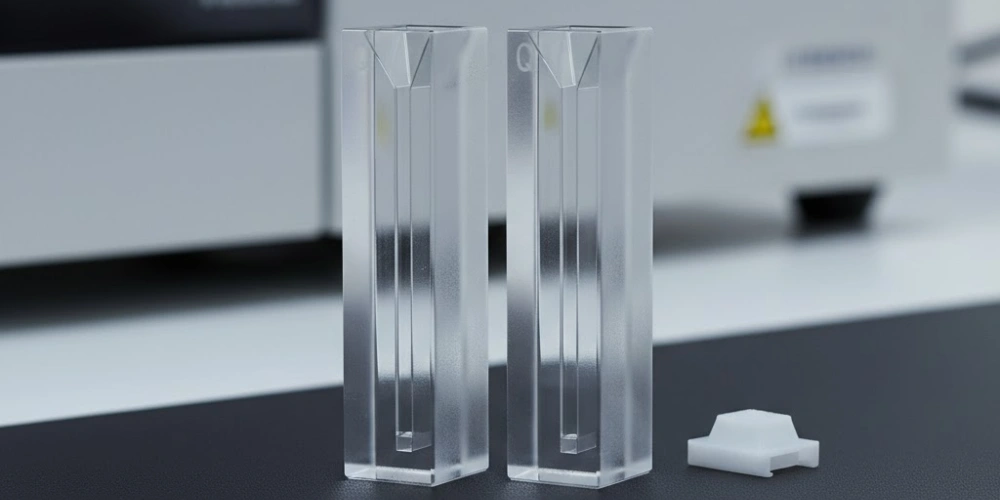
Choix de la longueur de trajet optique dans les cuvettes en quartz pour la spectroscopie UV-Vis et son incidence sur la précision des mesures
Au-delà de la transparence du matériau, la longueur de trajet optique d'une cuvette en quartz pour UV-Vis est le paramètre géométrique le plus déterminant, qui influe à la fois sur la précision et sur la plage dynamique linéaire des mesures d'absorbance.
La loi de Beer-Lambert et la dépendance linéaire par rapport à la longueur du trajet
La loi de Beer-Lambert exprime la relation fondamentale entre l'absorbance, la concentration de l'échantillon et la longueur du trajet optique : A = ε × c × l, où A est l'absorbance (sans dimension), ε est le coefficient d'atténuation molaire (L mol⁻¹ cm⁻¹), c est la concentration molaire (mol L⁻¹) et l est la longueur de trajet (cm). Cette loi prédit une relation strictement linéaire entre l’absorbance et la concentration pour une longueur de trajet fixe, ainsi qu’entre l’absorbance et la longueur de trajet pour une concentration fixe — mais cette linéarité ne vaut que dans une plage définie.
Les écarts par rapport à la linéarité de Beer-Lambert deviennent significatifs lorsque les valeurs d'absorbance dépassent environ 1,5 AU (ce qui correspond à une transmittance inférieure à 3,21 TP3T). À des absorbances élevées, la lumière parasite à l'intérieur du spectrophotomètre — c'est-à-dire le rayonnement qui atteint le détecteur sans traverser l'échantillon — représente une fraction proportionnellement plus importante du signal détecté, ce qui entraîne un plafonnement de l'absorbance apparente en dessous de sa valeur réelle. De plus, à des concentrations élevées de soluté, les interactions intermoléculaires entre les espèces absorbantes modifient le coefficient d’atténuation molaire effectif, introduisant des écarts chimiques par rapport à la linéarité. Ces deux effets conduisent systématiquement à une sous-estimation de la concentration réelle, avec des erreurs pouvant atteindre 5 à 151 TP3T pour des valeurs d'absorbance supérieures à 2,0 AU..
La réduction de la longueur de trajet optique est une méthode physiquement précise permettant de rétablir la linéarité pour les échantillons concentrés. En divisant par deux la longueur de trajet optique, qui passe ainsi de 10 mm à 5 mm, on divise par deux l'absorbance mesurée à concentration constante, ce qui ramène les mesures dans la plage linéaire sans nécessiter de dilution de l'échantillon — un avantage essentiel lorsque les volumes d'échantillon sont limités ou lorsque la dilution risquerait d'altérer les équilibres de la solution.
Longueurs de trajet standard dans les cuvettes en quartz et leurs applications correspondantes
Les fabricants produisent des cuvettes en quartz pour le spectrophotogramme UV-Vis dans une plage de longueurs de trajet optique allant d'environ trois ordres de grandeur, de 0,1 mm à 100 mm, afin de couvrir toute la gamme des concentrations d'échantillons rencontrées dans la pratique analytique.
Cuvettes à courte longueur de trajet optique — 0,1 mm, 0,2 mm et 0,5 mm — sont utilisés pour les échantillons à forte concentration, tels que les solutions-mères de protéines non diluées, les solutions concentrées de colorants et les formulations pharmaceutiques aux concentrations de process. Avec une longueur de trajet optique de 0,1 mm, un échantillon présentant un coefficient d'atténuation molaire de 10 000 L mol⁻¹ cm⁻¹ reste dans la plage linéaire jusqu'à des concentrations d'environ 150 mg/mL pour une protéine globulaire type — une plage inaccessible avec des cuvettes standard de 10 mm. La Longueur de trajet de 10 mm La cuvette est la norme universelle pour les mesures UV-Vis de routine ; elle offre une plage d'absorbance utile d'environ 0,05 à 1,5 AU pour la plupart des échantillons de laboratoire. Les cuvettes à grande longueur de parcours de 20 mm, 50 mm et 100 mm étendre la sensibilité aux échantillons à concentrations infimes, notamment les échantillons d'eau environnementale analysés pour détecter des contaminants aromatiques, les étalons d'impuretés pharmaceutiques ultra-dilués et les espèces chromophores à faible concentration dans le cadre de la recherche écologique.
Cuvettes en quartz micro et submicro pour l'analyse UV-Vis de petits volumes
Les contraintes liées au volume des échantillons, particulièrement courantes en génomique, en protéomique et en biologie unicellulaire, ont conduit au développement de formats de cuvettes en quartz micro et submicro qui conservent toutes leurs performances optiques tout en nécessitant des volumes nettement plus faibles.
Une cuvette en quartz standard d'une longueur de trajet optique de 10 mm nécessite environ 3,0 à 3,5 ml d'échantillon pour remplir la chambre optique située au-dessus du trajet du faisceau. Les cuvettes semi-micro réduisent cette quantité à 1,4 à 1,7 ml en réduisant la largeur de la chambre interne tout en conservant une longueur de trajet de 10 mm. Les microcuvettes permettent de réduire encore davantage le volume requis à 0,6 à 0,7 ml, et les formats submicroniques permettent d'obtenir des mesures exploitables avec aussi peu que 70 µL, obtenu grâce à la conception d'une chambre interne extrêmement étroite (généralement de 3 mm × 3 mm de section transversale) dont la dimension Z — c'est-à-dire la hauteur du centre du faisceau au-dessus du fond de la cuvette — est définie avec précision et adaptée à la géométrie optique du spectrophotomètre.
La dimension Z est un paramètre de compatibilité essentiel: la plupart des spectrophotomètres UV-Vis de paillasse positionnent le centre du faisceau à 8,5 mm au-dessus de la base de la cuvette, tandis que certains appareils utilisent des dimensions Z de 15 mm ou 20 mm. Une incompatibilité entre la dimension Z de la cuvette et la hauteur du faisceau de l'instrument entraîne un décalage partiel du faisceau par rapport au volume de l'échantillon, ce qui produit des mesures d'absorbance anormalement faibles, impossibles à distinguer des signaux authentiques de faible concentration sans vérification indépendante.
Référence relative à la longueur de trajet optique et au volume des cuvettes en quartz
| Longueur du trajet optique (mm) | Volume typique de l'échantillon (mL) | Application primaire |
|---|---|---|
| 0.1 | 0.05-0.15 | Concentrés protéiques hautement concentrés, solutions d'API |
| 0.5 | 0,10–0,30 | Extraits biologiques concentrés |
| 1 | 0,40–0,70 | Échantillons de concentration intermédiaire |
| 10 | 15 h 00 – 15 h 50 | Mesures UV-Vis de routine universelles |
| 20 | 18 h 00 – 19 h 00 | Diluer les échantillons environnementaux |
| 50 | 15,0–17,0 | Analyse des contaminants à l'état de traces |
| 100 | 30,0–35,0 | Mesures de concentrations à l'échelle des ultra-traces |
Variations de conception géométrique des cuvettes en quartz destinées aux montages expérimentaux UV-Vis
Les spécifications relatives à la longueur de trajet optique et au volume concernent ce qui se passe le long de l'axe optique principal ; toutefois, la géométrie du corps de la cuvette elle-même introduit des variables supplémentaires qui affectent la qualité de la mesure dans certaines configurations expérimentales spécifiques.
Cuvettes polies sur les quatre faces par rapport aux cuvettes polies sur les deux faces
Les cuvettes polies des deux côtés présentent des surfaces optiquement planes et exemptes de rayures uniquement sur les deux faces perpendiculaires au faisceau d'excitation — c'est-à-dire les faces que le rayonnement de mesure traverse effectivement. Les deux faces latérales restantes sont polies pour obtenir une finition mate, ce qui est suffisant pour garantir la stabilité mécanique et faciliter la manipulation, mais ne convient pas d'un point de vue optique pour la transmission du rayonnement. Les cuvettes à polissage double face conviennent parfaitement aux mesures d'absorbance standard. où le rayonnement pénètre par une face polie et ressort par la face polie opposée, en suivant un seul axe optique linéaire.
Les cuvettes polies sur les quatre faces présentent la même finition optique sur chacune de leurs quatre faces verticales, ce qui permet au rayonnement d'entrer ou de sortir par n'importe quelle face sans pertes par diffusion. Cette configuration est indispensable pour spectroscopie de fluorescence, où le rayonnement d'excitation pénètre par une face et où la fluorescence émise est collectée à 90° par une face adjacente. Cela est également nécessaire pour spectroscopie de dichroïsme circulaire (CD), où la géométrie d'interaction dépend d'un contrôle précis de la polarisation grâce à des surfaces optiquement homogènes, ainsi que pour les mesures de rotation optique. Le surcoût de fabrication lié au polissage de quatre faces au lieu de deux — ce qui représente généralement un 30–50% premium par rapport à des spécifications équivalentes de polissage bilatéral — n’est justifiée que lorsque la géométrie de mesure nécessite un accès optique latéral.
Une méthode d'identification pratique: les cuvettes polies sur deux faces présentent généralement une opacité visible ou un aspect givré sur leurs parois latérales lorsqu'on les observe sous un éclairage oblique, tandis que les cuvettes polies sur les quatre faces apparaissent uniformément transparentes sous tous les angles.
Cuvettes en quartz à parois noires et suppression de la lumière parasite dans les mesures de fluorescence
Les cuvettes standard à quatre faces polies, bien qu'optiquement accessibles sous tous les angles, introduisent un artefact spécifique dans les mesures de fluorescence : le rayonnement d'excitation réfléchi par les parois internes de la cuvette atteint le détecteur d'émission, ce qui entraîne la superposition d'un signal de fond sur le spectre de fluorescence réel. Cet artefact est particulièrement marqué aux longueurs d'onde d'émission proches de la longueur d'onde d'excitation — dans la région du pic de diffusion Raman de l'eau et dans la partie initiale de la bande d'émission de fluorescence.
Les cuvettes en quartz à parois noires permettent de remédier à cet artefact en appliquant un revêtement noir opaque sur les deux faces latérales et la face arrière, ne laissant transparentes et polies que la face avant (entrée d'excitation) et la face d'émission à 90°. Le revêtement noir absorbe le rayonnement d'excitation réfléchi et diffusé avant qu'il n'atteigne le détecteur d'émission, ce qui réduit le bruit de fond dû à la lumière parasite de facteurs compris entre 10 et 100× par rapport aux cuvettes standard à quatre parois polies utilisées dans les expériences de fluorescence. Le corps en quartz d'une cuvette à parois noires reste en silice fondue de qualité UV ; seul le revêtement de la surface externe diffère.
En spectroscopie de fluorescence, l'utilisation d'une cuvette standard à parois transparentes plutôt que d'une cuvette à parois noires a pour conséquence pratique un signal de fond plus intense et structuré sur le plan spectral. ce qui réduit la sensibilité, fausse les spectres d'émission aux longueurs d'onde situées dans une plage d'environ 30 à 50 nm autour de la longueur d'onde d'excitation, et compromet la précision quantitative pour les échantillons à faible fluorescence.
Variantes de conception des cuvettes en quartz et leurs applications
| Type de conception | Visages polis | Contrôle de la lumière parasite | Application primaire |
|---|---|---|---|
| Polissage des deux côtés | 2 (recto et verso) | Standard | Absorbance UV-Vis |
| Polissage sur les quatre côtés | 4 (toutes verticales) | Standard | Fluorescence, spectroscopie CD |
| À parois noires | 2 (face avant + face d'émission) | Améliorée | Fluorescence avec un faible bruit de fond |
| Circulation continue | 2 ou 4 | Standard | Détecteurs HPLC à débit continu |
| Cylindrique | Courbe continue | Limitée | Dichroïsme circulaire spécialisé |
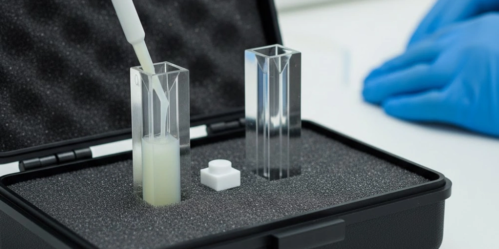
Profils de résistance chimique des cuvettes en quartz pour spectrométrie UV-Vis selon les matrices d'échantillons
La supériorité spectroscopique du quartz de silice fondue s'accompagne d'un avantage tout aussi important en termes de résistance chimique — un avantage qui détermine la gamme d'échantillons qu'une cuvette peut accueillir en toute sécurité sans que ses surfaces optiques ne subissent de dégradation.
Compatibilité de la silice fondue avec les acides et les solvants organiques
La résistance chimique de la silice fondue aux acides et aux solvants organiques découle directement de la stabilité thermodynamique du réseau de liaisons Si-O. L'énergie de dissociation de la liaison Si-O, qui est d'environ 452 kJ/mol est supérieure à celle de la plupart des liaisons métal-oxygène présentes dans les verres à plusieurs composants, ce qui explique pourquoi la silice fondue résiste aux réactifs qui attaquent facilement la verrerie de laboratoire classique.
Les acides minéraux forts — notamment l'acide chlorhydrique à toutes les concentrations, l'acide sulfurique jusqu'à une concentration d'environ 70%, l'acide nitrique et l'acide phosphorique à température ambiante — n'attaquent pas de manière mesurable les surfaces en silice fondue pendant les durées d'exposition habituelles en laboratoire. Les tampons acides aqueux couramment utilisés en biochimie (tampons citrate, acétate, phosphate et MES à un pH compris entre 3 et 6) sont tout aussi inoffensifs. Les solvants organiques présentent une plage de compatibilité tout aussi large : éthanol, méthanol, isopropanol, acétone, acétonitrile, diméthylsulfoxyde (DMSO), dichlorométhane, chloroforme, toluène et tétrahydrofurane (THF) sont tous compatibles avec les cuvettes en quartz de silice fondue sans provoquer de gonflement, de lixiviation ni de dégradation optique de la surface.
Une distinction pratique essentielle Le quartz de silice fondue se distingue des matériaux polymères utilisés pour les cuvettes : les solvants organiques qui provoquent immédiatement l'apparition de fissures, un voile ou la dissolution des cuvettes en PMMA ou en polystyrène — en particulier les solvants chlorés, les cétones et les hydrocarbures aromatiques — sont tolérés sans conséquence par le quartz, ce qui fait des cuvettes en quartz de silice fondue le seul choix pratique pour les mesures UV-Vis dans des systèmes de solvants non aqueux.
L'exception cruciale concernant l'acide fluorhydrique et les alcalis concentrés
Malgré sa grande compatibilité chimique, le quartz de silice fondue présente deux faiblesses chimiques bien définies dont il faut tenir compte sans exception dans la pratique de laboratoire.
L'acide fluorhydrique (HF) réagit avec le SiO₂ selon la réaction stœchiométrique suivante : SiO₂ + 4HF → SiF₄↑ + 2H₂O, produisant des composés volatils tétrafluorure de silicium2 et de l'eau. Cette réaction se produit à des vitesses mesurables même à des concentrations de HF aussi faibles que 0,1% et à température ambiante, provoquant la corrosion des surfaces optiques polies des cuvettes en quartz en quelques minutes à peine après le contact. La corrosion de surface entraîne des pertes par diffusion permanentes et une distorsion du front d'onde qui ne peuvent être éliminées par un nettoyage ; Une cuvette en quartz ayant été exposée à l'acide fluorhydrique doit être considérée comme irrémédiablement endommagée et mise hors service.. L'hydroxyde de sodium concentré (NaOH > 30%) et l'hydroxyde de potassium (KOH > 20%) attaquent la silice fondue selon un mécanisme de dissolution plus lent : les ions hydroxyde rompent les liaisons Si-O-Si par hydrolyse, avec des vitesses de dissolution d'environ 0,1 à 1 µm par heure à température ambiante, en présence d'un alcali concentré. L'acide phosphorique concentré, à des températures supérieures à 100 °C, attaque également la silice fondue à des vitesses qui augmentent fortement avec la température.
Pour les échantillons nécessitant de l'acide fluorhydrique, un alcali concentré ou de l'acide phosphorique concentré chaud, les récipients alternatifs appropriés sont ceux en polytétrafluoroéthylène (PTFE) ou en perfluoroalcoxy (PFA), dont aucun ne réagit avec ces réactifs.
Compatibilité chimique des cuvettes en quartz de silice fondue
| Catégorie de réactifs | Exemple | Compatibilité | Notes |
|---|---|---|---|
| Acides minéraux dilués | HCl, H₂SO₄, HNO₃ | ✓ Compatible | Toutes les concentrations, à température ambiante |
| Solvants organiques | EtOH, acétone, DMSO, CHCl₃ | ✓ Compatible | Pas de gonflement ni de dégradation optique |
| Tampons aqueux (pH 3–9) | PBS, HEPES, citrate | ✓ Compatible | Plage de pH biologique standard |
| Alcalis dilués (< 5%) | NaOH, KOH | ⚠ Attention | Exposition lente ; réduire au minimum le temps d'exposition |
| Alcalis concentrés | NaOH > 30% | ✗ Incompatible | Dissolution en surface en quelques heures |
| Acide fluorhydrique | HF (toute concentration) | ✗ Incompatible | Gravure immédiate et irréversible |
| H₃PO₄ concentré chaud | > 85%, > 100 °C | ✗ Incompatible | Accélération thermique de l'attaque par le SiO₂ |
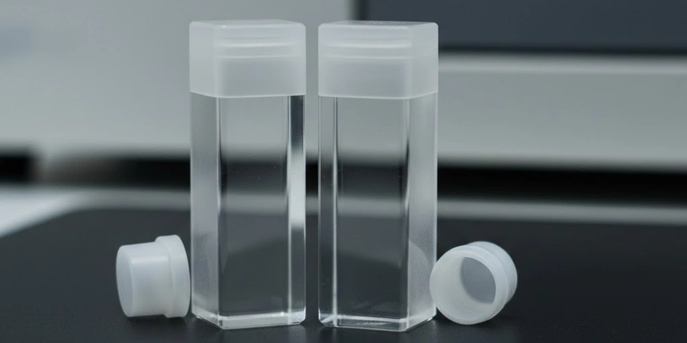
Pourquoi les cuvettes en verre ne donnent pas de résultats satisfaisants lors des mesures UV-Vis en dessous de 320 nm
Les cuvettes en verre restent omniprésentes dans les laboratoires d'enseignement et en colorimétrie dans le domaine du visible ; cependant, leurs limites spectroscopiques dans le domaine de l'ultraviolet sont suffisamment importantes pour les rendre scientifiquement inadaptées à une grande partie des applications analytiques.
Composition chimique du verre borosilicaté et du verre sodocalcique
Les cuvettes de laboratoire à usage commercial sont fabriquées soit en verre borosilicaté, soit en verre sodocalcique — deux compositions de silicates à plusieurs composants qui contiennent des proportions importantes d'agents de formation de réseau et de modificateurs autres que la silice.
Verre borosilicaté (comme par exemple le Schott DURAN et le Corning Pyrex) contient environ 81% de SiO₂, 13% de B₂O₃, 4% de Na₂O et 2% d'Al₂O₃ en poids. Le trioxyde de bore (B₂O₃) est ajouté pour réduire le coefficient de dilatation thermique, ce qui améliore la résistance aux chocs thermiques ; toutefois, les motifs structuraux bore-oxygène introduisent des transitions électroniques dans le domaine des UV qui sont absentes du SiO₂ pur. Verre sodocalcique contient généralement 72% de SiO₂, 14% de Na₂O, 9% de CaO et 5% de MgO — une composition optimisée pour la maniabilité et le coût plutôt que pour les performances optiques. Les oxydes modificateurs de réseau (Na₂O, CaO, MgO) perturbent le réseau Si-O, créant ainsi des sites d'oxygène non pontants qui forment des centres de défauts absorbant les UV.
Des traces d'impuretés métalliques dans les deux types de verre — en particulier les ions Fe²⁺ et Fe³⁺ présents à des concentrations comprises entre 50 et 200 ppm dans le verre optique standard — génèrent des bandes d'absorption UV intenses. Le Fe³⁺ produit des bandes d'absorption vers 225 nm et 320 nm grâce à transitions d-d liées au champ de ligands3 et les transitions par transfert de charge, tandis que le Fe²⁺ contribue à l'absorption vers 200 nm. Même à des concentrations inférieures à 100 ppm sur une longueur de trajet de 10 mm, ces bandes d’absorption du fer produisent des contributions d’absorbance comprises entre 0,1 et 0,5 AU à 280 nm — des valeurs qui masquent les signaux des échantillons biologiques dilués.
Le seuil d'absorption des UV du verre et ses conséquences spectroscopiques
Le seuil d'absorption dans l'ultraviolet d'un matériau de cuvette est généralement défini comme la longueur d'onde à laquelle l'absorbance intrinsèque du matériau est égale à 1,0 UA pour une longueur de trajet de 10 mm — point à partir duquel seule une partie de 10% du rayonnement incident est transmise et où le rapport signal/bruit s'est dégradé à un niveau qui rend la mesure quantitative peu fiable.
Pour le verre borosilicaté, cette longueur d'onde de coupure se situe entre 290 et 320 nm en fonction de la composition spécifique du verre et de sa teneur en fer. Pour le verre sodocalcique, la limite se situe généralement à 320–350 nm. Ces limites impliquent que les cuvettes en verre borosilicaté ne peuvent pas être utilisées pour des mesures à 280 nm (quantification des protéines par absorbance des composés aromatiques), à 260 nm (quantification des acides nucléiques), 254 nm (surveillance de la stérilisation par UV) et entre 214 et 220 nm (absorbance des liaisons peptidiques et quantification des protéines à faible longueur d’onde). Lorsqu’une cuvette en verre est utilisée pour un dosage protéique à 280 nm, le verre lui-même contribue à une absorbance d’environ 0,3–0,8 UA qui varie d'une cuvette à l'autre, alors que leurs caractéristiques sont en principe identiques — une erreur systématique liée au lot qui ne peut être corrigée par une simple mesure à blanc.
Les conséquences spectroscopiques s'enchaînent: une dérive de la ligne de base due aux variations de l'absorbance du verre en fonction de la température, des pics d'absorption apparents aux longueurs d'onde correspondant aux limites d'absorption du verre (qui peuvent être attribués à tort aux chromophores de l'échantillon), et une plage dynamique linéaire réduite qui contraint toute quantification à se situer dans la partie supérieure, non linéaire, de la courbe de Beer-Lambert.
Artefacts d'autofluorescence et de diffusion dans le verre aux longueurs d'onde UV
Au-delà de l'absorption directe, les cuvettes en verre génèrent des artefacts optiques secondaires dans le domaine des UV qui compromettent encore davantage la précision des mesures.
Autofluorescence du verre — l'émission spontanée de rayonnement visible suite à une excitation par rayons UV — résulte de transitions électroniques au sein de centres de défauts structurels et de contaminants organiques incorporés lors de la fabrication du verre. Lorsque le verre borosilicaté est irradié à 280 nm, il émet une fluorescence à large bande dont le pic se situe près de 400 à 450 nm, avec des rendements quantiques qui varient d'un lot de verre à l'autre. Dans un spectrophotomètre UV-Vis standard à faisceau unique, cette fluorescence contribue au signal détecté aux longueurs d’onde où la bande passante du monochromateur chevauche le spectre d’émission, ce qui entraîne une réduction apparente de l’absorbance de l’échantillon — un artefact qui varie de manière non linéaire avec l’intensité d’excitation et qui est absent des mesures à blanc réalisées avec le solvant seul dans une cuvette en quartz.
Les inclusions microscopiques présentes dans le verre — bulles de gaz piégées, zones de fusion non homogénées et précipités cristallins — agissent comme Centres de diffusion de Mie pour le rayonnement UV. La diffusion de Mie provenant de particules sphériques dont le diamètre est comparable à la longueur d’onde de mesure (100 à 300 nm pour le rayonnement UV) génère un bruit de fond dépendant de la longueur d’onde qui augmente fortement vers les longueurs d’onde plus courtes, imitant ainsi le profil d’absorption des particules colloïdales. En pratique, une cuvette en verre utilisée pour des mesures à 220 nm peut présenter une contribution apparente à l’absorbance due à la diffusion qui dépasse 0,5 UA — supérieure à l'absorbance de l'échantillon authentique pour de nombreux échantillons biologiques dilués.
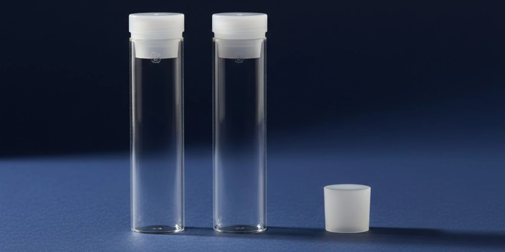
Comparaison des performances de transmission entre des cuvettes en verre de quartz, en plastique et en saphir
Pour choisir le matériau de cuvette approprié, il est nécessaire de procéder à une comparaison systématique des quatre catégories de matériaux à la disposition des spectroscopistes de laboratoire — quartz de silice fondue, verre borosilicaté, polyméthacrylate de méthyle (PMMA) et saphir — en fonction des exigences spécifiques des mesures UV-Vis.
Quartz de silice fondue Il présente la fenêtre de transmission la plus large en pratique, la meilleure compatibilité chimique et la meilleure stabilité optique de surface parmi les quatre matériaux, pour un coût de fabrication unitaire proportionnellement plus élevé. Sa transmission de 170 nm à 2 700 nm, avec une absorbance de base inférieure à 0,01 AU à 260 nm, en fait le matériau de référence par rapport auquel tous les autres sont évalués.
Verre borosilicaté Il offre une transmission comparable à celle du quartz au-delà de 320 nm et dans l'ensemble du spectre visible (400–800 nm), ce qui le rend adapté — et économique — aux dosages colorimétriques, à la cinétique enzymatique suivie aux longueurs d'onde du spectre visible, ainsi qu'à toute mesure ne nécessitant pas d'accès aux UV en dessous de 320 nm. Sa coupure UV située entre 290 et 320 nm et sa sensibilité à l’autofluorescence sous irradiation UV le rendent inadapté à la région du proche-UV.
PMMA et polystyrène Les cuvettes sont des formats à usage unique, jetables, dont les longueurs d'onde de coupure UV sont de 300–320 nm pour le PMMA et 340 à 360 nm pour le polystyrène — des limites qui les restreignent à la colorimétrie dans le spectre visible. Leurs principaux avantages sont leur prix et leur praticité : ils éliminent les risques de contamination croisée dans les environnements cliniques et à haut débit où l’utilisation de protocoles jetables est obligatoire. Les solvants organiques dissolvent ou fissurent immédiatement les cuvettes en plastique, et leurs surfaces non optiques présentent une diffusion importante. Saphir (Al₂O₃) les cuvettes transmettent à partir d'environ de 145 nm à 5 500 nm dotés d’une résistance chimique et d’une dureté mécanique exceptionnelles (9 sur l’échelle de Mohs), ce qui les rend techniquement supérieurs à la silice fondue pour les applications dans l’ultraviolet sous vide en dessous de 160 nm. Cependant, la biréfringence du saphir — due à sa structure cristalline trigonale — complique les mesures polarimétriques, et la difficulté de sa fabrication limite son utilisation à des applications de recherche spécialisées.
Comparaison de la transmission et de la compatibilité des matériaux des cuvettes
| Propriété | Quartz de silice fondue | Verre borosilicaté | Plastique PMMA | Saphir |
|---|---|---|---|---|
| Plage de transmission (nm) | 170–2 700 | 320–2 500 | 300–900 | 145–5 500 |
| Seuil de coupure UV à 1,0 UA / 10 mm (nm) | < 175 | 290–320 | 300–320 | < 150 |
| Utilisable en dessous de 260 nm | ✓ Oui | ✗ Non | ✗ Non | ✓ Oui |
| Résistance aux solvants organiques | Excellent | Bon | Pauvre | Excellent |
| Résistance HF | ✗ Non | ✗ Non | ✓ Oui | ✓ Oui |
| Autofluorescence sous rayonnement UV | Négligeable | Important | Modéré | Négligeable |
| Dureté de surface (échelle de Mohs) | 7 | 6-7 | 3 | 9 |
| Réutilisable | ✓ Oui | ✓ Oui | ✗ Non | ✓ Oui |
| Birefringence | Aucun | Aucun | Aucun | Présent |
Erreurs de mesure courantes liées au matériau ou à l'état de la cuvette
Même lorsque le matériau de cuvette approprié a été choisi, les erreurs de mesure imputables à l'état de la cuvette — rayures, contamination résiduelle, mauvaise orientation et bulles emprisonnées — constituent une source persistante de problèmes liés à la qualité des données en spectroscopie UV-Vis.
-
Dérive de la ligne de base et absorbance à blanc non nulle sont les artefacts liés aux cuvettes les plus fréquemment rencontrés. Lorsqu’une cuvette en quartz présentant une contamination de surface est utilisée pour une mesure à blanc, la ligne de base enregistrée intègre l’absorbance du contaminant comme étant nulle, ce qui entraîne une sous-estimation de la même valeur de l’absorbance réelle pour toutes les mesures d’échantillons suivantes. Un film protéique présent sur la surface optique d’une cuvette en quartz peut contribuer 0,05 à 0,2 UA d'absorbance apparente à 280 nm — une erreur suffisante pour entraîner une surestimation de la concentration en protéines de l'ordre de 10 à 50% dans un dosage standard de Bradford ou un dosage UV direct. En revanche, la dérive de la ligne de base induite par la température provient de l'échantillon plutôt que de la cuvette : l'indice de réfraction des solutions aqueuses varie d'environ −0,0001 par °C, ce qui modifie les pertes par réflexion de Fresnel aux interfaces de la cuvette et entraîne une dérive lente de l'absorbance qui se caractérise par sa réversibilité une fois la température stabilisée.
-
Valeurs d'absorbance anormalement élevées qui ne correspondent pas aux concentrations connues des échantillons proviennent souvent d'une diffusion induite par des rayures sur la surface optique de la cuvette plutôt que d'une absorption élevée de l'échantillon. Une simple rayure traversant la section transversale du faisceau peut augmenter l'absorbance apparente de 0,05–0,5 UA, en fonction de la profondeur et de la largeur de la rayure, la contribution due à la diffusion augmentant fortement aux longueurs d'onde plus courtes. Pour distinguer la diffusion due à la rayure de l'absorption réelle de l'échantillon, il faut mesurer la cuvette présentant une anomalie apparente par rapport à une cuvette de référence propre en utilisant la même solution témoin ; la diffusion due à la rayure se traduira par un décalage persistant de la ligne de base, tandis que l'absorption réelle de l'échantillon varie en fonction de la concentration de celui-ci.
-
Faible reproductibilité des mesures — un coefficient de variation supérieur à 1–2% entre les mesures répétées d'échantillons identiques — est souvent dû à une orientation irrégulière de la cuvette lors de son insertion. La plupart des cuvettes ne sont pas parfaitement carrées : les variations d'épaisseur des parois de ±0,01-0,05 mm La présence de faces opposées modifie la longueur de trajet effective selon la face exposée au faisceau. Le fait d'adopter une orientation d'insertion constante (marquée à l'aide d'un feutre de laboratoire ou par alignement sur le repère d'orientation du fabricant) permet généralement de réduire la variabilité de l'absorbance liée à l'orientation à une valeur inférieure à 0,31 TP3T.
-
Artéfacts de bulles provoquent des pics d’absorbance soudains et importants — dépassant souvent 1,0 AU — à des longueurs d’onde qui, autrement, se comportent normalement. Une bulle occupant ne serait-ce qu’une fraction de la section transversale du faisceau réfléchit pratiquement tout le rayonnement incident loin du détecteur, simulant ainsi une absorption quasi totale de l’échantillon. Ces bulles proviennent du gaz dissous qui s'échappe de la solution lorsque les échantillons sont transférés de la chambre froide à température ambiante, de l'introduction turbulente de l'échantillon à l'aide de pipettes à passage étroit, ainsi que des résidus de solvant de rinçage piégés dans des cuvettes mal séchées. Chauffage progressif à température ambiante avant la mesure, introduction lente de l'échantillon le long de la paroi de la cuvette plutôt que directement dans le faisceau, et séchage minutieux entre chaque utilisation empêchent de manière fiable la formation de bulles.
Vérification de l'intégrité d'une cuvette en quartz UV-Vis avant chaque série de mesures
La mise en place d'une brève procédure de vérification avant d'utiliser une cuvette en quartz pour des mesures UV-Vis quantitatives permet d'éviter l'accumulation d'erreurs systématiques non corrigées dans les ensembles de données expérimentales.
-
Vérification de la transmission de référence C'est le contrôle préalable à la mesure le plus révélateur. Remplir la cuvette d’eau de qualité HPLC (ou du solvant pur qui sera utilisé dans l’expérience) et effectuer un balayage de 190 nm à 350 nm par rapport à une référence à l’air permet de détecter à la fois la contamination résiduelle (absorbance élevée à des longueurs d’onde caractéristiques) et la diffusion de surface (ligne de base élevée qui s’élève uniformément vers les longueurs d’onde plus courtes). Une cuvette propre en quartz de silice fondue de qualité UV, remplie d’eau de qualité HPLC, devrait présenter une absorbance inférieure à 0,05 AU à 200 nm, moins de 0,02 AU à 230 nm et moins de 0,01 AU à 260 nm par rapport à un blanc d'air dans les conditions standard du spectrophotomètre. Tout écart supérieur à ces seuils indique soit une contamination résiduelle (nécessitant un nettoyage supplémentaire), soit un endommagement de la surface optique (nécessitant le remplacement de la cuvette).
-
Inspection visuelle sous un éclairage oblique complète le contrôle de base par spectrophotométrie en mettant en évidence les rayures, les opacités et les éclats mécaniques qui provoquent une diffusion sans pour autant produire de pics d'absorption spectrale distinctifs. En tenant la cuvette à environ 30° par rapport à un tube fluorescent ou à une source de lumière à fibre optique et en examinant les faces optiques en lumière transmise, les rayures apparaissent sous forme de stries linéaires brillantes ; l’opacité se présente comme une lueur diffuse à l’intérieur du corps en verre ; les éclats mécaniques apparaissent comme des zones brillantes aux contours nets au niveau des coins ou des bords de la cuvette. Toute cuvette présentant des rayures à l’intérieur de la zone centrale 80% de la face optique — la zone traversée par le faisceau du spectrophotomètre — doit être mise hors service pour les mesures quantitatives.
-
Vérification par paires appariées Cette procédure est nécessaire lorsque l'on utilise des spectrophotomètres à double faisceau avec des cuvettes d'échantillon et de référence distinctes. Le fait de remplir les deux cuvettes avec une solution témoin identique et de mesurer l'absorbance de l'une par rapport à l'autre dans la plage de 200 à 400 nm permet de quantifier leur équivalence photométrique. Une paire appariée doit présenter des différences d'absorbance inférieures à 0,005 UA sur l'ensemble de la gamme de longueurs d'onde; les paires dont le nombre dépasse Différence de transmittance de 0,51 TP3T pour toute longueur d'onde comprise dans la plage de mesure doivent être réajustés ou remplacés, car ce décalage introduit une erreur de ligne de base dépendante de la longueur d'onde qui ne peut pas être annulée par une simple mesure à blanc.
-
Critères de remplacement Pour les cuvettes en quartz UV-Vis, la durée de vie est déterminée par leurs performances optiques plutôt que par leur âge chronologique ou leur nombre d'utilisations. Une cuvette qui satisfait au test de transmission de référence et à l'inspection visuelle continue de fournir des mesures fiables, quelle que soit la durée de son utilisation. À l'inverse, une cuvette qui échoue au test de référence malgré un nettoyage minutieux — présentant une absorbance élevée persistante supérieure à 0,05 AU à 260 nm dans un solvant de qualité HPLC — présente une dégradation irréversible de sa surface optique et ne doit plus être utilisé pour les analyses quantitatives UV-Vis.
Conclusion
Le choix du matériau des cuvettes UV-Vis a des conséquences directes sur l'intégrité des données sur l'ensemble du spectre. Le quartz de silice fondue se distingue du verre, du plastique et de la plupart des matériaux concurrents car sa structure moléculaire — un réseau continu de SiO₂ présentant une bande interdite de 8,9 eV — transmet les longueurs d’onde comprises entre 170 nm et 2 700 nm sans absorption, d’autofluorescence ni de dégradation de surface en présence d’acides et de solvants organiques. Les cuvettes en verre présentent des défaillances en dessous de 320 nm en raison d’impuretés de métaux de transition, de défauts structurels et de compositions d’oxydes à plusieurs composants qui entraînent une absorption UV, une dérive de la ligne de base et des artefacts de fluorescence. Le choix d’une cuvette adaptée, en fonction de la longueur de trajet optique, de la géométrie et des exigences de nettoyage, n’est pas un aspect secondaire de la spectroscopie UV-Vis : c’est le fondement physique sur lequel repose chaque résultat quantitatif.
FAQ
Une cuvette en verre peut-elle être utilisée pour n'importe quelle mesure UV-Vis ?
Les cuvettes en verre peuvent être utilisées pour des mesures effectuées exclusivement au-dessus de 320 nm : colorimétrie dans le domaine du visible, dosages de cinétique enzymatique suivis entre 400 et 800 nm, et mesures de turbidité par absorbance. Elles ne conviennent pas aux mesures nécessitant des longueurs d’onde inférieures à 320 nm, notamment la quantification des protéines à 280 nm, la quantification des acides nucléiques à 260 nm, ou tout dosage reposant sur l’absorbance des liaisons aromatiques ou peptidiques dans le proche ultraviolet.
Quelle cuvette en quartz à quelle longueur de trajet optique est la plus couramment utilisée pour la plupart des applications UV-Vis ?
La cuvette en quartz de 10 mm de longueur de trajet optique constitue la norme universelle, car elle offre une plage d’absorbance utile d’environ 0,05 à 1,5 AU pour les concentrations d’échantillons typiques de la plupart des analyses biologiques et chimiques, correspond directement à la longueur de trajet supposée dans les valeurs tabulées des coefficients d'atténuation molaire (qui sont traditionnellement exprimées en L mol⁻¹ cm⁻¹, où 1 cm = 10 mm), et est compatible avec la géométrie optique de pratiquement tous les spectrophotomètres de paillasse disponibles dans le commerce.
À quelle fréquence faut-il remplacer une cuvette en quartz ?
La fréquence de remplacement est déterminée par les performances optiques, et non par la durée d'utilisation. Une cuvette en quartz qui satisfait à un test de transmission de la ligne de base — présentant une absorbance inférieure à 0,05 AU à 260 nm dans un solvant de qualité HPLC — et qui ne présente aucune rayure sur la surface optique centrale peut rester en service indéfiniment. Son remplacement est indiqué lorsqu’une absorbance de la ligne de base élevée et persistante, supérieure à ce seuil, persiste malgré un nettoyage approfondi, ce qui confirme des dommages irréversibles à la surface.
Existe-t-il un matériau de cuvette plus performant que le quartz pour les analyses UV-Vis ?
Le saphir (Al₂O₃) présente une fenêtre de transmission plus large que la silice fondue, s'étendant d'environ 145 nm dans l'ultraviolet sous vide à 5 500 nm dans l'infrarouge moyen. Toutefois, pour les applications UV-Vis en laboratoire limitées à la plage 190–800 nm, le quartz de silice fondue offre des performances équivalentes à celles du saphir tout en évitant la biréfringence inhérente à ce dernier — une propriété qui complique les mesures polarimétriques et de dichroïsme circulaire —, ce qui fait du quartz de silice fondue de qualité UV le choix pratique optimal pour la grande majorité des applications spectroscopiques UV-Vis.
Références :
-
La spectroscopie de dichroïsme circulaire mesure la différence d'absorption de la lumière polarisée circulairement à gauche et à droite par des molécules chirales ; elle nécessite des parois de cuvette optiquement homogènes afin de préserver l'intégrité de l'état de polarisation.↩
-
Le tétrafluorure de silicium est le produit gazeux volatil issu de la réaction entre l'acide fluorhydrique et le dioxyde de silicium ; sa formation est à l'origine de la gravure irréversible des surfaces en silice fondue lorsqu'elles sont exposées à l'HF.↩
-
Les transitions de champ de ligands sont des excitations électroniques au sein des ions de métaux de transition, provoquées par la dissociation des niveaux d'énergie des orbitales d sous l'effet du champ électrostatique des ligands environnants, ce qui donne lieu à des bandes d'absorption caractéristiques dans l'ultraviolet et le visible dans les verres contenant des métaux.↩