إن اختيار مادة غير مناسبة للكفيت يؤدي إلى تشويه البيانات الطيفية دون أن يلاحظ أحد — ولا يكتشف معظم الباحثين هذا الخطأ إلا بعد أن تصبح النتائج غير قابلة للتفسير.
لا تُعد مادة الكوفيت مسألة ثانوية في التحليل الطيفي بالأشعة فوق البنفسجية والمرئية (UV-Vis)؛ بل هي متغير أساسي يحدد بشكل مباشر ما إذا كانت قراءات الامتصاص تعكس التركيب الكيميائي للعينة أم أنها ناتجة عن تشوهات في الجهاز. تتناول هذه المقالة الخصائص البصرية والهيكلية والكيميائية والتشغيلية التي تجعل الكوارتز المادة المعيارية لقياسات الأشعة فوق البنفسجية والمرئية (UV-Vis)، مع إجراء مقارنة منهجية لأدائه مقارنةً بالبدائل المصنوعة من الزجاج والبلاستيك والياقوت عبر النطاق الطيفي الكامل.
يبدأ اختيار المواد في مجال التحليل الطيفي بفهم المتطلبات الفعلية للجهاز من الوعاء الذي يحمل العينة. فعندما يقوم مقياس الطيف الضوئي للأشعة فوق البنفسجية والمرئية (UV-Vis) بمسح الأطوال الموجية من 190 نانومتر إلى 800 نانومتر، يجب أن يقوم كل مكون بصري في مسار الضوء — بما في ذلك الكفيت — بنقل الإشعاع دون امتصاصه أو تشتيته أو إصدار فلورة. فالكفيتة التي تتداخل مع الحزمة الضوئية عند أي طول موجي ضمن هذا النطاق تتسبب في حدوث خطأ منهجي لا يمكن تصحيحه بالاعتماد على المعالجة البرمجية اللاحقة وحدها.
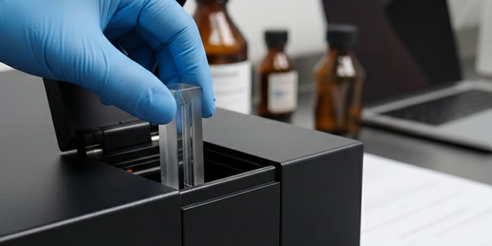
تحدد الشفافية البصرية للنافذة اختيار مادة الكفيت
تنقل كل مادة بصرية الإشعاع بشكل انتقائي، وتحدد حدود هذا الانتقائية ما إذا كانت الكفيتة صالحة علمياً للاستخدام في تجارب الأشعة فوق البنفسجية والمرئية (UV-Vis) أم أنها غير متوافقة معها من الناحية الأساسية.
ما هي نافذة النقل في المواد البصرية؟
تُعرف «نافذة النفاذية» بأنها نطاق الأطوال الموجية التي تسمح المادة خلاله بمرور الإشعاع الكهرومغناطيسي دون توهين ملحوظ. ينشأ التوهين عن ثلاث ظواهر متنافسة: الامتصاص، والانعكاس، والتشتت، حيث يساهم كل منها في فقدان الإشارة بطريقة تعتمد على الطول الموجي.
على المستوى الذري، يحدث الامتصاص عندما تتطابق طاقة الفوتون الساقط مع فجوة الطاقة بين الحالات الأساسية للإلكترونات والحالات المثارة داخل البنية الجزيئية أو البلورية للمادة. أما المواد ذات فجوات النطاقات الإلكترونية الواسعة — حيث تتجاوز الطاقة المطلوبة لترقية الإلكترونات إلى الحالات المثارة طاقة فوتونات الأشعة فوق البنفسجية — فتُظهر نوافذ نقل واسعة ذات أطوال موجية قصيرة. وعلى العكس من ذلك، فإن المواد التي تحتوي على أيونات المعادن الانتقالية، أو الهياكل العضوية المترافقة، أو العيوب الهيكلية، تمتلك حالات طاقة إلكترونية متوسطة تمتص الأشعة فوق البنفسجية بسهولة، مما يحجب بشكل فعال النفاذية تحت أطوال الموجات الفاصلة المميزة لها.
التأثير العملي على اختيار الكفيت مباشر: أي مادة يقع حافة امتصاصها ضمن النطاق الطيفي للقياس ستُركب منحنى الامتصاص الخاص بها على إشارة العينة، مما يجعل تصحيح خط الأساس بدقة أمراً مستحيلاً.
النطاق الطيفي للأشعة فوق البنفسجية والمرئية ومتطلباته البصرية الصارمة
يمتد النطاق الطيفي للأشعة فوق البنفسجية والمرئية (UV-Vis) عادةً من من 190 نانومتر إلى 800 نانومتر، والتي تنقسم بدورها إلى مناطق الأشعة فوق البنفسجية العميقة (190–280 نانومتر)، والأشعة فوق البنفسجية القريبة (280–400 نانومتر)، والضوء المرئي (400–800 نانومتر). وتفرض كل منطقة فرعية متطلبات محددة على شفافية مادة الكفيت.
تُعد منطقة الضوء المرئي (400–800 نانومتر) مرنة نسبيًا؛ حيث يمرر كل من الزجاج والكوارتز الضوء بشكل كافٍ عبر هذا النطاق، مما يجعل اختيار المادة أقل أهمية بالنسبة للفحوصات اللونية التي تُجرى حصريًّا في منطقة الضوء المرئي. أما النطاق القريب من الأشعة فوق البنفسجية (280–400 نانومتر)، فيبدأ في الكشف عن محدودية الزجاج ومعظم البوليمرات، حيث تتجاوز حواف امتصاصها النطاق الذي يبدأ من أقل من 320 نانومتر صعودًا. ويُعد النطاق العميق للأشعة فوق البنفسجية (190–280 نانومتر) هو الأكثر صعوبة: هناك أقل من ثلاث مواد عملية تجاريًا لصناعة الكفيتات — الكوارتز المصنوع من السيليكا المصهورة، والكوارتز الصناعي المخصص للأشعة فوق البنفسجية، والياقوت — تحافظ على شفافية كافية عند أطوال موجية أقل من 220 نانومتر.
يقع قياس كمية البروتين عند 280 نانومتر، وقياس كمية الحمض النووي عند 260 نانومتر، وامتصاص الرابطة الببتيدية عند 215 نانومتر، وتحديد خصائص الأحماض الأمينية العطرية في النطاق 250–290 نانومتر، كلها ضمن نطاق الأشعة فوق البنفسجية القريبة أو بالقرب منه. وبالنسبة لهذه التطبيقات، التي تمثل نسبة كبيرة من عمليات التحليل الطيفي الروتينية في المختبرات، شفافية مادة الكوفيت عند الطول الموجي الأقل من 320 نانومتر أمر لا يمكن التنازل عنه.
كيف تستفيد قياسات الأشعة فوق البنفسجية والمرئية من الخصائص البصرية لخلايا القياس الكوارتزية
الشفافية البصرية وحدها لا تفسر بشكل كامل السبب الذي يجعل قياسات الأشعة فوق البنفسجية والمرئية تستفيد بشكل كبير من كفيت كوارتز التصنيع. وتمتد هذه الميزة من البنية الجزيئية مروراً بدقة التصنيع وصولاً إلى جودة السطح.
البنية الجزيئية للسيليكا المدمجة وشفافيتها للأشعة فوق البنفسجية
السيليكا المذابة — وهي المادة الأساسية المستخدمة في كفيتات الكوارتز الخاصة بالأشعة فوق البنفسجية والمرئية — هي مادة صلبة غير متبلورة تتكون بالكامل من ثاني أكسيد السيليكون (SiO₂) المرتب في شبكة عشوائية متصلة من رباعيات الأوجه [SiO₄] التي تشترك في الزوايا. وعلى عكس الكوارتز البلوري، الذي يتميز بشبكة بلورية منظمة بعيدة المدى، تفتقر السيليكا المدمجة إلى الانتظام الهيكلي الدوري، مما يزيل الانكسار المزدوج ويجعله متجانسًا بصريًّا في جميع الاتجاهات.
تعد البنية الإلكترونية للرابطة Si-O عاملاً أساسياً في شفافية السيليكا المذابة للأشعة فوق البنفسجية. وتبلغ فجوة النطاق للسيليكا المذابة عالية النقاء حوالي 8.9 إلكترون فولت، وهو ما يتوافق مع بداية الامتصاص عند حوالي 140 نانومتر في منطقة الأشعة فوق البنفسجية الفراغية. تحمل الفوتونات عند 190 نانومتر طاقة تبلغ حوالي 6.5 إلكترون فولت — وهي أقل بكثير من العتبة المطلوبة لإثارة الإلكترونات عبر فجوة النطاق بين السيليكون والأكسجين — مما يعني أن الأشعة فوق البنفسجية ذات الأطوال الموجية ذات الصلة بالتحليل الطيفي المختبري تمر عبر السيليكا المنصهرة النقية دون امتصاص إلكتروني. ويشكل هذا تباينًا حادًا مع الزجاج متعدد المكونات، حيث تُحدث أكاسيد المُشبِّع حالات إلكترونية تحت فجوة النطاق تمتص الأشعة فوق البنفسجية عند أطوال موجية تصل إلى 350 نانومتر.
يُنتج السيليكا المدمجة الاصطناعية عن طريق التحلل المائي باللهب أو الترسيب الكيميائي بالبلازما (CVD) باستخدام SiCl₄، وتحقق تركيزات هيدروكسيل (OH) أقل من 1 جزء في المليون ومستويات شوائب معدنية أقل من 20 جزء في المليار، وكلاهما عنصران أساسيان للحفاظ على الشفافية للأشعة فوق البنفسجية. تحتفظ بلورات الكوارتز الطبيعية، حتى بعد تنقيتها، بآثار ضئيلة من الشوائب التي تسبب ظهور نطاقات امتصاص بالقرب من 245 نانومتر (مرتبطة بمراكز نقص الأكسجين) و214 نانومتر (مرتبطة بمراكز E')، مما يجعلها أقل كفاءة من السيليكا المصهورة الاصطناعية في تطبيقات الأشعة فوق البنفسجية العميقة.
نطاق النفاذية الضوئية لخزنة الكوارتز من الأشعة فوق البنفسجية العميقة إلى الأشعة تحت الحمراء القريبة
يُظهر السيليكا المصهور عالي النقاء نفاذية قابلة للقياس تبدأ من حوالي 170 نانومتر إلى 2,700 نانومتر، حيث تغطي نطاقًا طيفيًّا لا يمكن لأي مادة بصرية أخرى فعالة من حيث التكلفة أن تضاهيه بالكامل. وفي نطاق الأشعة فوق البنفسجية والمرئية (UV-Vis) على وجه التحديد (190–800 نانومتر)، تتجاوز قيم النفاذية عادةً في كفيتة من السيليكا المذابة من الدرجة المخصصة للأشعة فوق البنفسجية، التي يبلغ طول مسارها الضوئي 10 ملم، 85% عند 200 نانومتر و92% عند 250 نانومتر، حيث تُعزى الخسائر في المقام الأول إلى انعكاس فريسنل عند واجهتي التلامس بين الهواء والزجاج، وليس إلى الامتصاص الداخلي.
تتميز الدرجات التجارية الثلاث لكؤوس السيليكا المذابة بشكل أساسي بمحتواها من مجموعات الهيدروكسيل وما يرتبط بها من امتصاص للأشعة تحت الحمراء. السيليكا المنصهرة بالأشعة فوق البنفسجية (منخفضة المحتوى من OH، < 10 جزء في المليون من OH) تحقق نفاذية مثالية عند الطول الموجي الأقل من 250 نانومتر، مما يجعلها الخيار المناسب للتحليل الطيفي بالأشعة فوق البنفسجية العميقة. أما السيليكا المصهورة من الدرجة القياسية (عالية OH، 400–1,000 جزء في المليون من OH) فتُظهر انخفاضًا طفيفًا في نفاذية الأشعة فوق البنفسجية بسبب نغمات امتصاص OH بالقرب من 245 نانومتر، لكنها تقدم أداءً مقبولًا لمعظم تطبيقات الأشعة فوق البنفسجية القريبة فوق 220 نانومتر. أما السيليكا المصهورة من الدرجة IR، فهي تُحسّن النفاذية في النطاق 2,000–3,500 نانومتر على حساب بعض الأداء في نطاق الأشعة فوق البنفسجية.
بالنسبة لتحديد كميات الأحماض النووية والبروتينات — وهما أكثر تطبيقات الأشعة فوق البنفسجية والمرئية (UV-Vis) شيوعًا في المختبرات البيولوجية — توفر كفيتات السيليكا المذابة المخصصة للأشعة فوق البنفسجية، التي يبلغ طول مسار الضوء فيها 10 ملم، قيم امتصاص خط الأساس أقل من 0.01 وحدة امتصاص (AU) عند 260 نانومتر و280 نانومتر، مما يوفر هامش القياس اللازم لإجراء قياس كمي دقيق عبر نطاق تركيز واسع.
تجانس معامل الانكسار ونعومة السطح عبر درجات كفيتات الكوارتز المختلفة
يبلغ معامل الانكسار للسيليكا المذابة عند طول الموجة 589 نانومتر (خط الصوديوم D) حوالي 1.458، مع منحنى تشتت يتغير بشكل متوقع من 1.534 عند 193 نانومتر إلى 1.440 عند 1,064 نانومتر. والأهم من القيمة المطلقة لمعامل الانكسار هو تناسقه المكاني عبر النافذة البصرية للكفيت: تتميز قطع السيليكا المذابة عالية الجودة بتجانس في معامل الانكسار أفضل من ±5 × 10⁻⁶، مما يضمن أن تشوه مقدمة الموجة الناتج عن جدران الكفيت يكون ضئيلًا جدًّا مقارنةً بالدقة الضوئية للجهاز.
تحدد جودة تشطيب السطح بشكل مباشر خسائر التشتت عند جدران الكفيت. ويؤدي تلميع السيليكا المصهورة بالدرجة البصرية إلى تحقيق قيم خشونة سطح تقل عن 0.5 نانومتر RMS (الجذر التربيعي المتوسط)، مما يحافظ على خسائر التشتت السطحي أقل من 0.1% عبر نطاق الأشعة فوق البنفسجية والمرئية. ويتطلب التلميع وفقًا لهذه المواصفات عملية صقل متعددة المراحل باستخدام مواد كاشطة تزداد دقةً تدريجيًّا، تليها عملية تلميع بالقطران أو تشطيب مغناطيسي-ريولوجي — وهي عمليات خاصة بالتصنيع البصري وتختلف عن إنتاج الأواني الزجاجية المختبرية القياسية.
كويتات تلميع ذات وجهين — حيث لا يخضع للتشطيب البصري سوى الوجهين المتعامدين مع شعاع الضوء — وهي كافية لإجراء قياسات الامتصاص القياسية. كفيتات تلميع رباعية الجوانب، حيث يجب أن تكون جميع الأوجه الأربعة العمودية مزودة بنفس الطلاء البصري، وذلك من أجل إجراء التحليل الطيفي بالفلورة، الانحراف اللوني الدائري (CD)1، وقياسات الدوران البصري التي يدخل فيها الإشعاع أو يخرج من خلال الأوجه الجانبية.
ملخص الأداء البصري لخزانات الكوارتز
| المعلمة | السيليكا المنصهرة بالأشعة فوق البنفسجية | السيليكا المذابة القياسية | السيليكا المذابة من الدرجة IR |
|---|---|---|---|
| نطاق الإرسال (نانومتر) | 170–2,700 | 190–2,700 | 220–3,500 |
| محتوى OH (جزء في المليون) | < 10 | 400–1,000 | < 10 |
| النفاذية عند 200 نانومتر (%) | > 85 | 75–85 | 60–75 |
| النفاذية عند 260 نانومتر (%) | > 92 | > 90 | > 88 |
| معامل الانكسار عند 589 نانومتر | 1.458 | 1.458 | 1.458 |
| القيمة المتوسطة المربعة لخشونة السطح (نانومتر) | < 0.5 | < 0.5 | < 0.5 |
| الشوائب المعدنية (جزء في المليار) | < 20 | < 50 | < 20 |
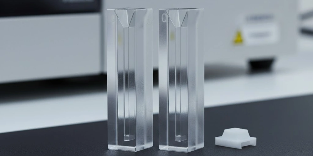
اختيار طول المسار في كفيتات الكوارتز للأشعة فوق البنفسجية والمرئية وتأثيره على دقة القياس
وبصرف النظر عن الشفافية المادية، فإن طول مسار الضوء في كفيتة الكوارتز المستخدمة في نطاق الأشعة فوق البنفسجية والمرئية (UV-Vis) هو المعلمة الهندسية الأكثر أهمية التي تؤثر على كل من دقة قياسات الامتصاص ونطاقها الديناميكي الخطي.
قانون بير-لامبرت والاعتماد الخطي على طول المسار
يعبر قانون بير-لامبرت عن العلاقة الأساسية بين الامتصاص وتركيز العينة وطول المسار البصري: أ = ε × ج × ل، حيث A هي الامتصاصية (بدون أبعاد)، وε هو معامل التوهين المولي (L mol⁻¹ cm⁻¹)، وc هو التركيز المولي (mol L⁻¹)، وl هو طول المسار (سم). يتنبأ هذا القانون بوجود علاقة خطية تمامًا بين الامتصاص والتركيز عند طول مسار ثابت، وبين الامتصاص وطول المسار عند تركيز ثابت — لكن هذه الخطية لا تسري إلا ضمن نطاق محدد.
تصبح الانحرافات عن خطية بير-لامبرت ملحوظة عندما تتجاوز قيم الامتصاص ما يقارب 1.5 وحدة أفريقية (ما يعادل نفاذية أقل من 3.2%). عند مستويات الامتصاص العالية، يشكل الضوء الشارد داخل جهاز قياس الطيف الضوئي — وهو الإشعاع الذي يصل إلى الكاشف دون المرور عبر العينة — نسبة أكبر نسبيًا من الإشارة المكتشفة، مما يتسبب في استقرار الامتصاص الظاهري عند مستوى أقل من قيمته الحقيقية. بالإضافة إلى ذلك، عند التركيزات العالية للمادة المذابة، تؤدي التفاعلات بين الجزيئات بين الأنواع الممتصة إلى تغيير معامل التوهين المولي الفعال، مما يؤدي إلى حدوث انحرافات كيميائية عن الخطية. يؤدي هذان العاملان إلى التقليل المنهجي من التقدير الحقيقي للتركيز، حيث تصل الأخطاء إلى 5–15% عند قيم الامتصاص التي تتجاوز 2.0 وحدة امتصاص (AU).
يُعد تقليل طول المسار طريقة دقيقة من الناحية الفيزيائية لاستعادة الخطية في العينات عالية التركيز. فخفض طول المسار إلى النصف — من 10 ملم إلى 5 ملم — يؤدي إلى خفض الامتصاصية المقاسة إلى النصف عند تركيز ثابت، مما يعيد القياسات إلى النطاق الخطي دون الحاجة إلى تخفيف العينة — وهي ميزة حاسمة عندما تكون أحجام العينات محدودة أو عندما يؤدي التخفيف إلى تغيير توازنات المحلول.
أطوال المسار القياسية عبر خلايا الكوارتز وتطبيقاتها المقابلة
تقوم الشركات المصنعة بإنتاج كفيتات كوارتز للأشعة فوق البنفسجية والمرئية (UV-Vis) بنطاق طول مسار يبلغ حوالي ثلاث مراتب من الحجم، من 0.1 ملم إلى 100 ملم، لتغطية التنوع الكامل لتركيزات العينات التي تُواجه في الممارسة التحليلية.
كفيتات ذات مسافة ضوئية قصيرة — 0.1 ملم، و0.2 ملم، و0.5 ملم — تُستخدم مع العينات عالية التركيز مثل محاليل البروتين غير المخففة، ومحاليل الأصباغ المركزة، والتركيبات الصيدلانية بتركيزات العملية. وعند طول مسار يبلغ 0.1 مم، تظل العينة التي يبلغ معامل التوهين المولي لها 10,000 لتر مول⁻¹ سم⁻¹ ضمن النطاق الخطي حتى تركيزات تصل إلى حوالي 150 ملغ/مل لبروتين كروي نموذجي — نطاق لا يمكن الوصول إليه باستخدام كفيتات قياسية مقاس 10 ملم. و طول المسار 10 ملم تُعد الكفيتة المعيار العالمي لقياسات الأشعة فوق البنفسجية والمرئية الروتينية، حيث توفر نطاق امتصاص عملي يتراوح بين 0.05 و1.5 وحدة امتصاص تقريبًا لمعظم العينات المختبرية. أما الكفيتات ذات مسار الضوء الطويل التي يبلغ طولها 20 ملم، و50 ملم، و100 ملم توسيع نطاق الحساسية ليشمل العينات ذات التركيزات النزرة، بما في ذلك عينات المياه البيئية التي يتم تحليلها للكشف عن الملوثات العطرية، ومعايير الشوائب الصيدلانية فائقة التخفيف، والمركبات الملونة ذات التركيزات المنخفضة في الأبحاث البيئية.
كفيتات كوارتز ذات حجم صغير وأصغر من الصغير لتحليل الأشعة فوق البنفسجية والمرئية (UV-Vis) لأحجام محدودة
دفعت القيود المتعلقة بحجم العينات — التي تنتشر بشكل خاص في مجالات الجينوميات والبروتينوميات وبيولوجيا الخلية الواحدة — إلى تطوير أنماط من الكفيتات الكوارتزية ذات الحجم الصغير وأصغر من الصغير، والتي تحافظ على الأداء البصري الكامل مع الحاجة إلى أحجام أصغر بكثير.
تتطلب كفيتة الكوارتز القياسية ذات طول المسار البالغ 10 ملم ما يقارب 3.0–3.5 مل من العينة لملء الحجرة البصرية فوق مسار الشعاع. وتقلل الكفيتات شبه الميكروية هذا المطلب إلى 1.4–1.7 مل عن طريق تضييق عرض الحجرة الداخلية مع الحفاظ على طول المسار البالغ 10 ملم. كما تقلل الكفيتات الدقيقة متطلبات الحجم إلى 0.6–0.7 مل، كما أن التنسيقات الأصغر من الميكرو توفر قياسات قابلة للاستخدام باستخدام ما لا يزيد عن 70 ميكرولتر، وقد تم تحقيق ذلك من خلال تصميم حجرة داخلية ضيقة للغاية (يبلغ مقطعها العرضي عادةً 3 مم × 3 مم) ذات بُعد Z محدد بدقة — وهو ارتفاع مركز الحزمة فوق قاعدة الكفيت — يتوافق مع الهندسة البصرية لجهاز قياس الطيف الضوئي.
يُعد البعد Z معلمة توافقية حاسمة: في معظم أجهزة قياس الطيف الضوئي للأشعة فوق البنفسجية والمرئية المكتبية، يتم وضع مركز الشعاع عند 8.5 مم فوق قاعدة الكوفيت، في حين تستخدم بعض الأجهزة أبعادًا على المحور Z تبلغ 15 مم أو 20 مم. ويؤدي عدم تطابق البعد Z للكوفيت مع ارتفاع شعاع الجهاز إلى عدم تغطية الشعاع لحجم العينة بشكل كامل، مما ينتج عنه قراءات امتصاص منخفضة بشكل غير طبيعي لا يمكن تمييزها عن الإشارات الحقيقية ذات التركيز المنخفض دون التحقق المستقل.
مرجع طول المسار والحجم لكأس الكوارتز
| طول المسار (مم) | حجم العينة النموذجي (مل) | التطبيق الأساسي |
|---|---|---|
| 0.1 | 0.05-0.15 | مخزونات بروتينية عالية التركيز، محاليل API |
| 0.5 | 0.10–0.30 | مستخلصات بيولوجية مركزة |
| 1 | 0.40–0.70 | عينات ذات تركيز متوسط |
| 10 | 3.00–3.50 | قياسات روتينية شاملة في نطاق الأشعة فوق البنفسجية والمرئية (UV-Vis) |
| 20 | 6.00–7.00 | تخفيف العينات البيئية |
| 50 | 15.0–17.0 | تحليل الملوثات النزرة |
| 100 | 30.0–35.0 | قياسات التركيزات الفائقة الصغرى |
الاختلافات في التصميم الهندسي لخلايا الكوارتز المستخدمة في أجهزة التجارب في نطاق الأشعة فوق البنفسجية والمرئية
تتناول مواصفات طول المسار والحجم ما يحدث على طول المحور البصري الرئيسي، إلا أن الشكل الهندسي لجسم الكفيت نفسه يُدخل متغيرات إضافية تؤثر على جودة القياس في تكوينات تجريبية محددة.
الكفيتات المصقولة من الجوانب الأربعة مقابل الكفيتات المصقولة من الجانبين
تتميز كفيتات التلميع ذات الوجهين بأسطح مستوية بصريًّا وخالية من الخدوش حصريًّا على الوجهين المتعامدين مع شعاع الإثارة — وهما الوجهان اللذان يمر عبرهما إشعاع القياس فعليًّا. أما الوجهان الجانبيان المتبقيان، فقد تم صقلهما ليصبحا غير لامعين، وهو ما يكفي لتحقيق الاستقرار الميكانيكي وسهولة المناولة، لكنه غير مناسب بصريًّا لنقل الإشعاع. تُعد كفيتات التلميع ذات الوجهين مناسبة تمامًا لقياسات الامتصاص القياسية حيث يدخل الإشعاع من أحد الأوجه المصقولة ويخرج من الوجه المصقول المقابل على طول محور بصري خطي واحد.
تتميز كفيتات التلميع ذات الجوانب الأربعة بنفس التشطيب البصري على جميع الجوانب الرأسية الأربعة، مما يسمح للإشعاع بالدخول أو الخروج عبر أي جانب دون حدوث خسائر ناتجة عن التشتت. ويُعد هذا التصميم ضروريًا لـ التحليل الطيفي الفلوري، حيث يدخل إشعاع الإثارة عبر أحد الأوجه ويتم جمع الفلورة المنبعثة بزاوية 90° عبر وجه مجاور. كما أنه ضروري أيضًا لـ التحليل الطيفي بالانحراف اللوني الدائري (CD)، حيث تعتمد هندسة التفاعل على التحكم الدقيق في الاستقطاب من خلال أسطح متجانسة بصريًّا، ولأغراض قياسات الدوران البصري. وتتمثل التكلفة الإضافية للتصنيع في صقل أربعة أوجه بدلاً من وجهين — وهو ما يمثل عادةً 30–50% ممتاز بالمقارنة مع المواصفات البولندية المكافئة ثنائية الجوانب — لا يكون ذلك مبرراً إلا عندما تتطلب هندسة القياس وصولاً بصرياً جانبيًّا.
طريقة عملية للتعرف على الهوية: عادةً ما تظهر الكفيتات المصقولة من الجانبين عتامةً مرئيةً أو مظهرًا بلوريًّا على جدرانها الجانبية عند النظر إليها تحت إضاءة مائلة، في حين تبدو الكفيتات المصقولة من الجوانب الأربعة شفافةً بشكل متجانس من جميع الزوايا.
أكواب الكوارتز ذات الجدران السوداء وقمع الضوء الشارد في قياسات الفلورة
تُحدث كفيتات التلميع القياسية ذات الجوانب الأربعة، على الرغم من إمكانية الوصول إليها بصريًّا من جميع الاتجاهات، تشويشًا معينًا في قياسات الفلورة: يصل إشعاع الإثارة المنعكس من جدران الكفيت الداخلية إلى كاشف الانبعاث، مما يؤدي إلى تراكب إشارة خلفية على طيف الفلورة الحقيقي. وتكون هذه الظاهرة الاصطناعية شديدة بشكل خاص عند أطوال موجات الانبعاث القريبة من طول موجة الإثارة — في منطقة ذروة تشتت رامان للماء وفي الجزء الأولي من نطاق انبعاث الفلورة.
تعالج أنابيب الكوارتز ذات الجدران السوداء هذه الظاهرة من خلال تطبيق طبقة سوداء غير شفافة على الوجهين الجانبيين والوجه الخلفي، بحيث لا يُترك سوى الوجه الأمامي (مدخل الإثارة) ووجه الانبعاث بزاوية 90 درجة شفافين ومصقولين. ويقوم الطلاء الأسود بامتصاص إشعاع الإثارة المنعكس والمشتت قبل أن يصل إلى كاشف الانبعاث، مما يقلل من خلفية الضوء الشارد بمقدار عوامل تتراوح بين 10 و100× مقارنةً بالكفيتات القياسية ذات الجوانب الأربعة المصقولة المستخدمة في تجارب الفلورة. ويظل جسم الكفيت ذو الجدران السوداء مصنوعًا من السيليكا المصهورة المخصصة للأشعة فوق البنفسجية؛ ولا يختلف سوى طلاء السطح الخارجي.
تتمثل النتيجة العملية لاستخدام كفيتة قياسية ذات جدران شفافة بدلاً من كفيتة ذات جدران سوداء في التحليل الطيفي الفلوري في ظهور إشارة خلفية مرتفعة وذات بنية طيفية وهو ما يقلل من الحساسية، ويشوه أطياف الانبعاث عند أطوال موجية تقع في نطاق 30–50 نانومتر تقريبًا من طول موجة الإثارة، ويؤثر سلبًا على الدقة الكمية بالنسبة للعينات ذات الفلورة الضعيفة.
أنواع تصميمات كفيتات الكوارتز وتطبيقاتها
| نوع التصميم | وجوه مصقولة | التحكم في الضوء الشارد | التطبيق الأساسي |
|---|---|---|---|
| ملمع على الوجهين | 2 (الأمام والخلف) | قياسي | الامتصاص في نطاق الأشعة فوق البنفسجية والمرئية |
| تلميع من الجوانب الأربعة | 4 (جميعها عمودية) | قياسي | الفلورة، التحليل الطيفي بالانعكاس الكروي (CD) |
| ذو الجدران السوداء | 2 (الجزء الأمامي + واجهة الانبعاث) | محسّن | الفلورة ذات الخلفية المنخفضة |
| التدفق المباشر | 2 أو 4 | قياسي | أجهزة الكشف في نظام HPLC، التدفق المستمر |
| أسطواني | منحنى متواصل | محدودة | الديكرويزم الدائري المتخصص |
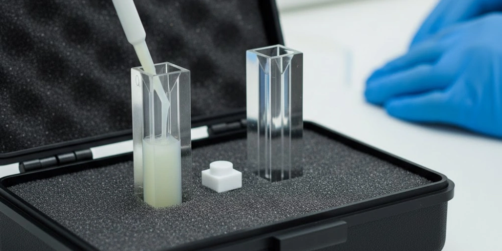
خصائص مقاومة المواد الكيميائية لخلايا الكوارتز الخاصة بالأشعة فوق البنفسجية والمرئية عبر مصفوفات العينات المختلفة
إن التفوق الطيفي للكوارتز المصنوع من السيليكا المذابة يقترن بميزة لا تقل أهمية تتمثل في المتانة الكيميائية — وهي الميزة التي تحدد نطاق العينات التي يمكن للكوفيت استيعابها بأمان دون أن تتعرض أسطحه البصرية للتلف.
توافق السيليكا المذابة مع الأحماض والمذيبات العضوية
تنبع مقاومة السيليكا المذابة الكيميائية للأحماض والمذيبات العضوية مباشرةً من الاستقرار الديناميكي الحراري لشبكة الروابط Si-O. طاقة تفكك الرابطة Si-O التي تبلغ حوالي 452 كيلوجول/مول وهي تفوق تلك الموجودة في معظم الروابط المعدنية-الأكسجينية في الزجاج متعدد المكونات، وهو ما يفسر قدرة السيليكا المذابة على مقاومة الكواشف التي تهاجم بسهولة الأواني الزجاجية المختبرية التقليدية.
الأحماض المعدنية القوية — بما في ذلك حمض الهيدروكلوريك بجميع تركيزاته، وحمض الكبريتيك حتى تركيز يبلغ حوالي 70%، وحمض النيتريك، وحمض الفوسفوريك في درجة حرارة الغرفة — لا تسبب تآكلًا ملحوظًا لأسطح السيليكا المصهورة خلال فترات التعرض المعتادة في المختبر. كما أن المحاليل الحمضية المائية المستخدمة بشكل روتيني في الكيمياء الحيوية (محاليل السيترات، والأسيتات، والفوسفات، وMES عند درجة حموضة تتراوح بين 3 و6) غير ضارة بالمثل. وتمتاز المذيبات العضوية بنطاق توافق واسع بنفس القدر: الإيثانول، والميثانول، والإيزوبروبانول، والأسيتون، والأسيتونيتريل، وثنائي ميثيل سلفوكسيد (DMSO)، وثنائي كلورو الميثان، والكلوروفورم، والتولوين، والتتراهيدروفوران (THF) وهي جميعها متوافقة مع كفيتات الكوارتز المصنوعة من السيليكا المدمجة دون التسبب في انتفاخ أو تسرب أو تدهور الخصائص البصرية للسطح.
تمييز عملي بالغ الأهمية يتميز الكوارتز المصنوع من السيليكا المذابة عن مواد الكفيتات البوليمرية: فالمذيبات العضوية التي تتسبب في حدوث تشققات فورية في كفيتات PMMA أو البوليستيرين، أو تعكرها، أو إذابتها — لا سيما المذيبات المكلورة، والكيتونات، والهيدروكربونات العطرية — لا يؤثر على الكوارتز، مما يجعل كفيتات الكوارتز المصنوعة من السيليكا المذابة الخيار العملي الوحيد لقياسات الأشعة فوق البنفسجية والمرئية (UV-Vis) في أنظمة المذيبات غير المائية.
الاستثناء الحرج لحمض الهيدروفلوريك والقلويات المركزة
على الرغم من التوافق الكيميائي الواسع للسيليكا المذابة، فإنها تعاني من نقطتي ضعف كيميائيتين محددتين بوضوح يجب مراعاتهما دون استثناء في الممارسات المختبرية.
يتفاعل حمض الهيدروفلوريك (HF) مع SiO₂ من خلال التفاعل المتكافئ SiO₂ + 4HF → SiF₄↑ 2H₂O، مما يؤدي إلى تقلبات رباعي فلوريد السيليكون2 والماء. وتستمر هذه التفاعل بمعدلات قابلة للقياس حتى عند تركيزات HF منخفضة تصل إلى 0.1% وفي درجة حرارة الغرفة، مما يؤدي إلى تآكل الأسطح البصرية المصقولة لخلايا الكوارتز في غضون دقائق من التلامس. ويؤدي تآكل السطح إلى خسائر تشتت دائمة وتشوه في جبهة الموجة لا يمكن إزالتها بالتنظيف؛ يجب اعتبار أي كفيتة كوارتز تعرضت لحمض الهيدروفلوريك تالفة بشكل لا يمكن إصلاحه، ويجب إيقاف استخدامها. يعمل هيدروكسيد الصوديوم المركز (NaOH > 30%) وهيدروكسيد البوتاسيوم (KOH > 20%) على تآكل السيليكا المصهورة من خلال آلية إذابة أبطأ — حيث تعمل أيونات الهيدروكسيد على قطع الروابط Si-O-Si عن طريق التحلل المائي، بمعدلات إذابة تبلغ تقريبًا 0.1–1 ميكرومتر في الساعة في درجة حرارة الغرفة في محلول قلوي مركّز. كما أن حمض الفوسفوريك المركّز عند درجات حرارة تزيد عن 100 درجة مئوية يتسبب في تآكل السيليكا المصهورة بمعدلات تزداد بشكل حاد مع ارتفاع درجة الحرارة.
بالنسبة للعينات التي تتطلب استخدام حمض الهيدروفلوريك أو القلويات المركزة أو حمض الفوسفوريك المركّز الساخن، فإن الأوعية البديلة المناسبة هي حاويات مصنوعة من البولي تترافلوروإيثيلين (PTFE) أو البيرفلوروألكوكسي (PFA)، ولا يتفاعل أي منهما مع هذه الكواشف.
التوافق الكيميائي لخلايا القياس المصنوعة من الكوارتز والسيليكا المذابة
| فئة الكواشف | مثال على ذلك | التوافق | الملاحظات |
|---|---|---|---|
| الأحماض المعدنية المخففة | HCl، H₂SO₄، HNO₃ | ✓ متوافق | جميع التركيزات، درجة حرارة الغرفة |
| المذيبات العضوية | الإيثانول، الأسيتون، ثنائي ميثيل سلفوكسيد، كلورو ميثان | ✓ متوافق | لا يحدث تورم أو تدهور بصري |
| محاليل عازلة مائية (pH 3–9) | PBS، HEPES، سيترات | ✓ متوافق | النطاق البيولوجي القياسي لقيمة الرقم الهيدروجيني |
| القلويات المخففة (< 5%) | NaOH، KOH | ⚠ تحذير | تعريض بطيء؛ تقليل مدة التعريض إلى أدنى حد |
| القلويات المركزة | NaOH > 30% | ✗ غير متوافق | ذوبان السطح في غضون ساعات |
| حمض الهيدروفلوريك | HF (أي تركيز) | ✗ غير متوافق | الحفر الفوري الذي لا رجعة فيه |
| حمض الفوسفوريك المركز الساخن (H₃PO₄) | > 85%، > 100 درجة مئوية | ✗ غير متوافق | التسريع الحراري لعملية تآكل ثاني أكسيد السيليكون |
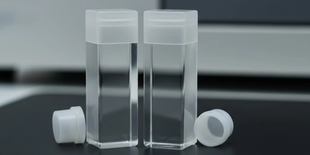
لماذا تعاني الكفيتات الزجاجية من قصور في قياسات الأشعة فوق البنفسجية والمرئية (UV-Vis) عند الطول الموجي الأقل من 320 نانومتر
لا تزال الكفيتات الزجاجية منتشرة على نطاق واسع في المختبرات التعليمية وفي قياس الألوان في النطاق المرئي، إلا أن قيودها الطيفية في منطقة الأشعة فوق البنفسجية شديدة بما يكفي لجعلها غير ملائمة علمياً لنسبة كبيرة من التطبيقات التحليلية.
التركيب الكيميائي للزجاج البورسليكاتي والزجاج الصودا-الجيري
يتم تصنيع الكفيتات الزجاجية المختبرية التجارية إما من زجاج البورسليكات أو زجاج الصودا والجير — وهما تركيبتان من السيليكات متعددتا المكونات تتضمنان نسبًا كبيرة من العناصر المكونة للشبكة غير السيليكا والمعدِّلات.
زجاج البورسليكات (مثل شوت دوران وكورنينغ بايركس) يحتوي على ما يقارب 81% من SiO₂، و13% من B₂O₃، و4% من Na₂O، و2% من Al₂O₃ بالوزن. يُضاف ثالث أكسيد البورون (B₂O₃) لتقليل معامل التمدد الحراري، مما يحسّن مقاومة الصدمات الحرارية، لكن الوحدات الهيكلية المكونة من البورون والأكسجين تُحدث انتقالات إلكترونية في نطاق الأشعة فوق البنفسجية، وهي انتقالات غير موجودة في SiO₂ النقي. زجاج الصودا والليمون يحتوي عادةً على 72% من SiO₂، و14% من Na₂O، و9% من CaO، و5% من MgO — تركيبة مُحسَّنة من حيث قابلية التشكيل والتكلفة بدلاً من الأداء البصري. تعمل أكاسيد مُعدِّلات الشبكة (Na₂O، CaO، MgO) على تعطيل شبكة Si-O، مما يؤدي إلى تكوين مواقع أكسجين غير متصلة تشكل مراكز عيوب تمتص الأشعة فوق البنفسجية.
الشوائب المعدنية النزرة في كلا النوعين من الزجاج — لا سيما أيونات Fe²⁺ وFe³⁺ الموجودة بتركيزات تتراوح بين 50 و200 جزء في المليون في الزجاج البصري القياسي — تولد نطاقات امتصاص قوية للأشعة فوق البنفسجية. ينتج Fe³⁺ نطاقات امتصاص بالقرب من 225 نانومتر و320 نانومتر من خلال انتقالات d-d في مجال الليجاند3 والانتقالات الناتجة عن نقل الشحنة، بينما يساهم Fe²⁺ في الامتصاص بالقرب من 200 نانومتر. وحتى عند تركيزات أقل من 100 جزء في المليون عبر مسار ضوئي يبلغ طوله 10 مم، تنتج نطاقات امتصاص الحديد هذه مساهمات في الامتصاص تتراوح بين 0.1 و0.5 وحدة امتصاص (AU) عند 280 نانومتر — وهي قيم تطغى على إشارات العينات البيولوجية المخففة.
حد امتصاص الأشعة فوق البنفسجية للزجاج وتبعاته الطيفية
يُعرَّف حد الامتصاص للأشعة فوق البنفسجية لمادة الكفيت تقليديًّا بأنه الطول الموجي الذي تساوي عنده السعة الامتصاصية الذاتية للمادة 1.0 وحدة فلكية عند طول مسار يبلغ 10 ملم — وهي النقطة التي لا يتم عندها تمرير سوى 10% من الإشعاع الساقط، وتكون نسبة الإشارة إلى الضوضاء قد تدهورت إلى مستوى يجعل القياس الكمي غير موثوق به.
بالنسبة للزجاج البورسليكاتي، يقع طول الموجة الفاصل هذا بين 290 و320 نانومتر اعتمادًا على التركيب المحدد للزجاج ومحتواه من الحديد. أما بالنسبة للزجاج الصودا-الكالسي، فإن الحد الفاصل عادةً ما يكون 320–350 نانومتر. وتعني هذه القيم الحدية أن كفيتات زجاج البورسليكات غير صالحة للاستخدام في القياسات عند 280 نانومتر (تحديد كمية البروتين عن طريق الامتصاص العطري)، و260 نانومتر (تحديد كمية الحمض النووي)، 254 نانومتر (مراقبة التعقيم بالأشعة فوق البنفسجية)، و214–220 نانومتر (امتصاص روابط الببتيد وقياس كمية البروتين عند الأطوال الموجية المنخفضة). وعند استخدام كفيت زجاجي في تحليل البروتين عند 280 نانومتر، يساهم الزجاج نفسه بامتصاص يبلغ تقريبًا 0.3–0.8 وحدة فلكية وهو ما يختلف بين الكوفيتات الفردية ذات المواصفات المتطابقة نظريًّا — وهو خطأ منهجي متغير بين الدُفعات لا يمكن تصحيحه بقياس فارغ واحد.
تتوالى النتائج الطيفية: انحراف خط الأساس الناجم عن التغيرات في امتصاص الزجاج المرتبطة بدرجة الحرارة، وظهور قمم امتصاص ظاهرية عند الأطوال الموجية المطابقة لحواف امتصاص الزجاج (والتي يمكن أن تُعزى خطأً إلى العوامل الملونة في العينة)، وتقلص النطاق الديناميكي الخطي الذي يُجبر جميع عمليات القياس الكمي على الانحصار في الجزء العلوي غير الخطي من منحنى بير-لامبرت.
الفلورة الذاتية والعيوب الناتجة عن التشتت في الزجاج عند أطوال الموجات فوق البنفسجية
بالإضافة إلى الامتصاص المباشر، تولد الكفيتات الزجاجية تشوهات بصرية ثانوية في نطاق الأشعة فوق البنفسجية، مما يقلل من دقة القياس بشكل أكبر.
الفلورة الذاتية للزجاج — الانبعاث التلقائي للإشعاع المرئي عقب الإثارة بالأشعة فوق البنفسجية — ينشأ عن انتقالات إلكترونية داخل مراكز العيوب الهيكلية والملوثات العضوية التي تتخلل الزجاج أثناء تصنيعه. وعندما يتعرض زجاج البورسليكات للإشعاع عند طول موجي يبلغ 280 نانومتر، فإنه يصدر فلورة واسعة النطاق تبلغ ذروتها بالقرب من 400–450 نانومتر، مع عوائد كمية تتفاوت بين دفعات الزجاج المختلفة. وفي مقياس الطيف الضوئي القياسي أحادي الشعاع للأشعة فوق البنفسجية والمرئية، يساهم هذا الفلوريسنس في الإشارة المكتشفة عند الأطوال الموجية التي يتداخل فيها نطاق مرور أحادي اللون مع طيف الانبعاث، مما يؤدي إلى انخفاض ظاهري في امتصاص العينة — وهو تشويه يتناسب بشكل غير خطي مع شدة الإثارة ويغيب عن قياسات العينة الفارغة التي تُجرى باستخدام المذيب وحده في كفيتة كوارتز.
تعمل الشوائب المجهرية في الزجاج — مثل فقاعات الغاز المحبوسة، ومناطق الصهر غير المختلطة، ورواسب البلورات الصغيرة — كـ مراكز تشتت مي بالنسبة للأشعة فوق البنفسجية. يؤدي تشتت مي الناتج عن الجسيمات الكروية التي يبلغ قطرها ما يعادل طول موجة القياس (100–300 نانومتر للأشعة فوق البنفسجية) إلى توليد خلفية تعتمد على طول الموجة وترتفع بشكل حاد مع انخفاض طول الموجة، مما يحاكي منحنى الامتصاص الخاص بالجسيمات الغروية. في الممارسة العملية، قد تُظهر الكفيتة الزجاجية المستخدمة في القياسات عند 220 نانومتر مساهمة امتصاصية ظاهرية ناتجة عن التشتت تتجاوز 0.5 وحدة فلكية — أكبر من قيمة الامتصاص في العينة الأصلية بالنسبة للعديد من العينات البيولوجية المخففة.
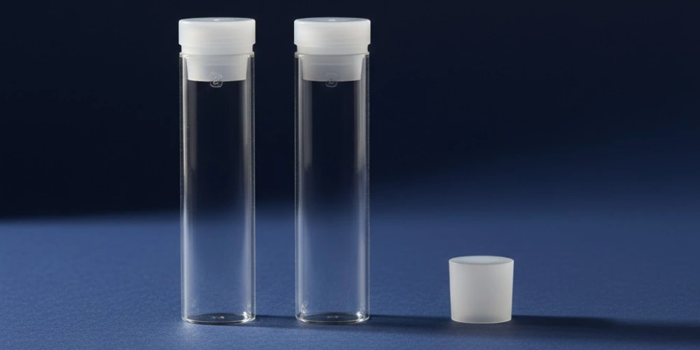
مقارنة أداء النقل بين أنابيب القياس المصنوعة من الزجاج الكوارتزي والبلاستيك والياقوت
يتطلب اختيار مادة الكفيت المناسبة إجراء مقارنة منهجية بين جميع فئات المواد الأربع المتاحة لأخصائيي الطيف الضوئي في المختبرات — الكوارتز المصنوع من السيليكا المصهورة، والزجاج البورسليكاتي، وبولي ميثيل ميثاكريلات (PMMA)، والياقوت — مع تقييمها وفقًا للمتطلبات المحددة لقياسات الأشعة فوق البنفسجية والمرئية.
الكوارتز المصنوع من السيليكا المذابة تتميز هذه المادة بأوسع نطاق عملي للانتقال الضوئي، وأعلى مستوى من التوافق الكيميائي، وأفضل استقرار بصري للسطح مقارنةً بالمواد الأربع الأخرى، مع تكلفة تصنيع أعلى للوحدة تبعًا لذلك. إن نطاق نفاذيتها الذي يمتد من 170 نانومتر إلى 2,700 نانومتر مع امتصاص أساسي أقل من 0.01 AU عند 260 نانومتر يجعلها المادة المرجعية التي تُقاس جميع المواد الأخرى على أساسها.
زجاج البورسليكات يحقق هذا المادة نفاذية مماثلة لنفاذية الكوارتز فوق 320 نانومتر وفي النطاق المرئي بأكمله (400–800 نانومتر)، مما يجعله مناسبًا — وفعالًا من حيث التكلفة — للاختبارات اللونية، وحركيات الإنزيمات التي تُراقب عند الأطوال الموجية المرئية، وأي قياس لا يتطلب التعرض للأشعة فوق البنفسجية تحت 320 نانومتر. إن حد قطع الأشعة فوق البنفسجية عند حوالي 290–320 نانومتر وقابليته للتألق الذاتي تحت إشعاع الأشعة فوق البنفسجية تجعله غير مناسب لمنطقة الأشعة فوق البنفسجية القريبة.
البلاستيك من نوع PMMA والبوليستيرين الكفيتات هي أدوات تستخدم لمرة واحدة وقابلة للتخلص منها، وتتميز بأطوال موجية لقطع الأشعة فوق البنفسجية تبلغ 300–320 نانومتر بالنسبة لـ PMMA و 340–360 نانومتر للبوليستيرين — القيود التي تحصر استخدامها في قياس الألوان ضمن النطاق المرئي. وتتمثل مزاياها الرئيسية في السعر وسهولة الاستخدام: فهي تقضي على مخاوف التلوث المتبادل في البيئات السريرية وبيئات الإنتاج عالي السعة التي يُشترط فيها اتباع بروتوكولات الاستخدام لمرة واحدة. فالمذيبات العضوية تؤدي إلى إذابة أو تشقق الكفيتات البلاستيكية على الفور، كما أن أسطحها غير المخصصة للاستخدامات البصرية تتسبب في تشتت كبير للضوء. الياقوت (ال₂O₃) تنقل الكفيتات من حوالي 145 نانومتر إلى 5,500 نانومتر التي تتميز بمقاومة كيميائية استثنائية وصلابة ميكانيكية (9 على مقياس موس)، مما يجعلها متفوقة تقنيًّا على السيليكا المصهورة في تطبيقات الأشعة فوق البنفسجية الفراغية التي تقل طول موجتها عن 160 نانومتر. ومع ذلك، فإن انكسار الضوء المزدوج في الياقوت — الناجم عن بنيته البلورية الثلاثية الزوايا — يعقّد القياسات الاستقطابية، كما أن صعوبة تصنيعه تقصر استخدامه على تطبيقات بحثية متخصصة.
مقارنة بين نفاذية المواد المستخدمة في الكفيتات وتوافقها
| الممتلكات | الكوارتز المصنوع من السيليكا المذابة | زجاج البورسليكات | بلاستيك PMMA | الياقوت |
|---|---|---|---|---|
| نطاق الإرسال (نانومتر) | 170–2,700 | 320–2,500 | 300–900 | 145–5,500 |
| حد قطع الأشعة فوق البنفسجية عند 1.0 وحدة فلكية / 10 ملم (نانومتر) | < 175 | 290–320 | 300–320 | < 150 |
| يمكن استخدامه عند طول موجي أقل من 260 نانومتر | ✓ نعم | ✗ لا | ✗ لا | ✓ نعم |
| مقاومة المذيبات العضوية | ممتاز | جيد | فقير | ممتاز |
| مقاومة التردد العالي جداً | ✗ لا | ✗ لا | ✓ نعم | ✓ نعم |
| الفلورة الذاتية تحت الأشعة فوق البنفسجية | ضئيل | مهم | معتدل | ضئيل |
| صلابة السطح (موس) | 7 | 6-7 | 3 | 9 |
| قابلة لإعادة الاستخدام | ✓ نعم | ✓ نعم | ✗ لا | ✓ نعم |
| الانكسار البيريفيرينجي | لا يوجد | لا يوجد | لا يوجد | حاضر |
أخطاء القياس الشائعة التي تعزى إلى مادة الكوفيت أو حالته
وحتى عند اختيار المادة المناسبة للكيويتة، فإن أخطاء القياس التي تُعزى إلى حالة الكيويتة — مثل الخدوش، والتلوث المتبقي، وسوء التوجيه، والفقاعات المحبوسة — تمثل مصدرًا مستمرًا لمشاكل جودة البيانات في التحليل الطيفي بالأشعة فوق البنفسجية والمرئية (UV-Vis).
-
انحراف خط الأساس وامتصاص الفراغ غير الصفري هي أكثر الأخطاء المرتبطة بالكوفيت شيوعًا. فعندما تُستخدم كوفيت كوارتز تراكمت على سطحها ملوثات لإجراء قياس فارغ، فإن خط الأساس المسجل يضم امتصاص الملوثات باعتباره صفرًا، مما يؤدي إلى أن جميع قياسات العينات اللاحقة تُقلل من قيمة الامتصاص الحقيقي بنفس المقدار. ويمكن أن يسهم وجود طبقة بروتينية على السطح البصري لكوفيت الكوارتز في 0.05–0.2 وحدة فلكية من الامتصاص الظاهري عند 280 نانومتر — وهو خطأ يكفي لإحداث انحراف في تقدير تركيز البروتين بمقدار 10–50% في اختبار برادفورد القياسي أو الاختبار المباشر بالأشعة فوق البنفسجية. وعلى النقيض من ذلك، فإن الانحراف في خط الأساس الناتج عن درجة الحرارة ينشأ من العينة نفسها وليس من الكفيت: حيث يتغير معامل الانكسار للمحاليل المائية بمقدار تقريبًا −0.0001 لكل درجة مئوية، مما يؤدي إلى تغيير خسائر الانعكاس الفريسنلي عند واجهات الكفيت، وينتج عن ذلك انحراف بطيء في الامتصاصية يتميز بقدرته على العودة إلى حالته الأصلية عند استقرار درجة الحرارة.
-
قراءات امتصاص مرتفعة بشكل غير معتاد التي لا تتوافق مع تركيزات العينات المعروفة غالبًا ما تنشأ عن التشتت الناتج عن الخدوش على السطح البصري للكوفيت، وليس عن ارتفاع امتصاص العينة. فوجود خدش واحد عبر المقطع العرضي للحزمة الضوئية يمكن أن يزيد الامتصاص الظاهري بمقدار 0.05–0.5 وحدة فلكية، اعتمادًا على عمق الخدش وعرضه، مع ارتفاع مساهمة التشتت بشكل حاد عند الأطوال الموجية الأقصر. ويتطلب التمييز بين تشتت الخدش والامتصاص الحقيقي للعينة قياس الكفيت الذي يبدو شاذًا مقارنةً بكفيت مرجعي نظيف باستخدام نفس المحلول الفارغ؛ حيث سيظل تشتت الخدش على شكل انحراف مستمر في خط الأساس، في حين يتغير الامتصاص الحقيقي للعينة تبعًا لتركيز العينة.
-
ضعف قابلية تكرار القياس — تجاوز معامل التباين 1–2% عبر القياسات المتكررة لعينات متطابقة — غالبًا ما يُعزى ذلك إلى عدم اتساق اتجاه إدخال الكفيت. فمعظم الكفيتات ليست مربعة تمامًا: حيث توجد تباينات في سماكة الجدران تبلغ ± 0.01-0.05 مم يؤدي التبديل بين الأوجه المتقابلة إلى تغيير طول المسار الفعال اعتمادًا على الوجه الذي يتعرض للشعاع. وعادةً ما يؤدي تحديد اتجاه إدخال ثابت (يُحدد بقلم مختبري أو عن طريق محاذاة علامة الاتجاه التي يحددها المصنع) إلى تقليل تباين الامتصاص المرتبط بالاتجاه إلى ما دون 0.3%.
-
الآثار الناتجة عن الفقاعات تؤدي إلى حدوث ارتفاعات مفاجئة وكبيرة في الامتصاص — غالبًا ما تتجاوز 1.0 وحدة امتصاص (AU) — عند أطوال موجية تكون سلوكية بشكل جيد في الأحوال العادية. فالفقاعة التي تمتد حتى لو كانت تغطي جزءًا ضئيلًا من المقطع العرضي للحزمة الضوئية تعكس عمليًّا كل الإشعاع الساقط بعيدًا عن الكاشف، مما يحاكي امتصاصًا شبه كامل من جانب العينة. وتنشأ الفقاعات من الغاز المذاب الذي يخرج من المحلول عند نقل العينات من التخزين البارد إلى درجة حرارة الغرفة، ومن إدخال العينة بشكل مضطرب عبر ماصات ذات فتحة ضيقة، ومن بقايا مذيب الشطف المحبوس في الكفيتات غير المجففة جيدًا. تسخين العينة برفق حتى تصل إلى درجة حرارة الغرفة قبل إجراء القياس، وإدخال العينة ببطء على طول جدار الكوفيت بدلاً من إدخالها مباشرةً في مسار الشعاع، والتجفيف التام بين الاستخدامات تمنع بشكل موثوق تكوّن الفقاعات.
التحقق من سلامة كفيتة الكوارتز الخاصة بالأشعة فوق البنفسجية والمرئية قبل كل عملية قياس
إن وضع إجراء تحقق موجز قبل استخدام كفيتة الكوارتز الخاصة بالأشعة فوق البنفسجية والمرئية (UV-Vis) في القياسات الكمية يمنع تراكم الأخطاء المنهجية غير المصححة عبر مجموعات البيانات التجريبية.
-
التحقق من معدل الانتقال الأساسي هو الفحص التمهيدي الأكثر إفادة قبل إجراء القياس. إن ملء الكفيت بماء من فئة HPLC (أو المذيب النقي الذي سيُستخدم في التجربة) وإجراء المسح من 190 نانومتر إلى 350 نانومتر مقابل مرجع الهواء يكشف عن كل من التلوث المتبقي (ارتفاع الامتصاصية عند أطوال موجية مميزة) والتشتت السطحي (ارتفاع خط الأساس الذي يرتفع بشكل منتظم نحو الأطوال الموجية الأقصر). يجب أن يُظهر الكفيت النظيف المصنوع من الكوارتز السيليكا المصهور من الدرجة المخصصة للأشعة فوق البنفسجية والمملوء بماء من الدرجة المخصصة للكروماتوغرافيا السائلة عالية الضغط (HPLC) امتصاصًا أقل من 0.05 وحدة فلكية عند 200 نانومتر، وأقل من 0.02 وحدة فلكية عند 230 نانومتر، وأقل من 0.01 وحدة فلكية عند 260 نانومتر مقارنةً بعينة هواء فارغة في ظل الظروف القياسية لجهاز قياس الطيف الضوئي. وتشير الانحرافات التي تتجاوز هذه الحدود إما إلى وجود تلوث متبقي (مما يستلزم تنظيفًا إضافيًا) أو إلى تلف السطح البصري (مما يستلزم استبدال الكفيت).
-
الفحص البصري تحت إضاءة مائلة ويكمل هذا الفحص الفحص الأساسي بالطيف الضوئي من خلال الكشف عن أنماط الخدوش، والضبابية، والكسور الميكانيكية التي تسبب التشتت دون أن تنتج بالضرورة سمات امتصاص طيفية مميزة. عند إمساك الكفيت بزاوية 30 درجة تقريبًا بالنسبة لأنبوب فلوري أو مصدر ضوء من الألياف البصرية وفحص الأوجه البصرية في الضوء المنقول، تظهر الخدوش على شكل خطوط خطية ساطعة؛ ويظهر التغيم على شكل توهج منتشر داخل جسم الزجاج؛ وتظهر الشقوق الميكانيكية على شكل مناطق ساطعة ذات حواف حادة عند زوايا الكفيت أو حوافه. أي كفيت تظهر عليه خدوش داخل الجزء المركزي 80% من السطح البصري — المنطقة التي يمر بها شعاع جهاز القياس الطيفي — يجب إيقاف تشغيلها عند إجراء القياسات الكمية.
-
التحقق من الأزواج المتطابقة يُعد هذا الإجراء ضروريًا عند استخدام أجهزة قياس الطيف الضوئي ثنائية الحزمة مع كفيتات منفصلة للعينة والمعيار. ويؤدي ملء الكفيتتين بمحلول فارغ متطابق وقياس الامتصاصية لأحدهما مقارنةً بالآخر في النطاق الطيفي 200–400 نانومتر إلى تحديد مدى تكافؤهما الضوئي كمياً. وينبغي أن يُظهر الزوج المتطابق فروقاً في الامتصاصية أقل من 0.005 وحدة فلكية عبر نطاق الطول الموجي الكامل؛ الأزواج التي يتجاوز عددها 0.5% فرق النفاذية عند أي طول موجي ضمن نطاق القياس يجب إعادة مطابقتها أو استبدالها، لأن عدم المطابقة يؤدي إلى حدوث خطأ في خط الأساس يعتمد على الطول الموجي ولا يمكن إزالته عن طريق قياس فارغ واحد.
-
معايير الاستبدال يتم تحديد عمر كفيتات الكوارتز المستخدمة في قياس الأشعة فوق البنفسجية والمرئية (UV-Vis) بناءً على الأداء البصري وليس على العمر الزمني أو عدد مرات الاستخدام. فالكفيتة التي تجتاز اختبار النفاذية الأساسي والفحص البصري تظل تقدم قياسات موثوقة بغض النظر عن المدة التي قضتها في الخدمة. وعلى العكس من ذلك، فإن الكفيتة التي تفشل في اجتياز الاختبار الأساسي على الرغم من تنظيفها بدقة — وتُظهر امتصاصية مرتفعة ومستمرة أعلى من 0.05 وحدة فلكية عند 260 نانومتر في مذيب من النوع المستخدم في التحليل الكروماتوغرافي السائل عالي الأداء (HPLC) — وقد تعرض سطحه البصري لتدهور دائم، لذا ينبغي سحبه من الاستخدام في الأعمال الكمية التي تعتمد على الأشعة فوق البنفسجية والمرئية.
الخاتمة
يُعد اختيار المواد المستخدمة في كفيتات الأشعة فوق البنفسجية والمرئية قرارًا له آثار مباشرة على سلامة البيانات عبر النطاق الطيفي بأكمله. يتميز الكوارتز المصنوع من السيليكا المذابة عن الزجاج والبلاستيك ومعظم المواد المنافسة لأن تركيبته الجزيئية — وهي شبكة SiO₂ متصلة ذات فجوة طيفية إلكترونية تبلغ 8.9 إلكترون فولت — تسمح بمرور الضوء من 170 نانومتر إلى 2,700 نانومتر دون امتصاص، أو فلورة ذاتية، أو تدهور السطح في وجود الأحماض والمذيبات العضوية. تفشل الخلايا الزجاجية في النطاق دون 320 نانومتر بسبب شوائب المعادن الانتقالية، والعيوب الهيكلية، وتركيبات الأكاسيد متعددة المكونات التي تسبب امتصاص الأشعة فوق البنفسجية، وانحراف خط الأساس، والتشوهات الفلورية. إن الاختيار الصحيح للكوب، بما يتناسب مع طول المسار والهندسة ومتطلبات التنظيف، ليس أمراً ثانوياً في التحليل الطيفي بالأشعة فوق البنفسجية والمرئية — بل هو الأساس الفيزيائي الذي تستند إليه كل نتيجة كمية.
الأسئلة الشائعة
هل يمكن استخدام كفيتة زجاجية في أي قياسات للأشعة فوق البنفسجية والمرئية؟
يمكن استخدام الكفيتات الزجاجية في القياسات التي تُجرى بالكامل فوق 320 نانومتر — مثل قياس الألوان في النطاق المرئي، واختبارات حركية الإنزيمات التي تُراقب في النطاق 400–800 نانومتر، وقياسات العكارة القائمة على الامتصاص. وهي غير مناسبة لأي قياس يتطلب استخدام أطوال موجية أقل من 320 نانومتر، بما في ذلك تحديد كمية البروتين عند 280 نانومتر، وتحديد كمية الحمض النووي عند 260 نانومتر، أو أي اختبار يعتمد على امتصاص الروابط العطرية أو الببتيدية في منطقة الأشعة فوق البنفسجية القريبة.
ما هو طول مسار الضوء القياسي لكفيتة الكوارتز المستخدمة في معظم تطبيقات الأشعة فوق البنفسجية والمرئية؟
تُعد الكفيتة الكوارتزية ذات طول المسار البالغ 10 ملم المعيار العالمي لأنها توفر نطاق عمل عملي للامتصاص يتراوح بين 0.05 و1.5 وحدة امتصاص (AU) تقريبًا لتركيزات العينات النموذجية في معظم التحاليل البيولوجية والكيميائية، ويطابق بشكل مباشر طول المسار المفترض في قيم معامل التوهين المولي المجدولة (التي تُسجل عادةً بوحدة L mol⁻¹ cm⁻¹, حيث 1 سم = 10 مم)، كما أنه متوافق مع الهندسة البصرية لجميع أجهزة قياس الطيف الضوئي المكتبية التجارية تقريبًا.
كم مرة يجب استبدال كفيت الكوارتز؟
يتم تحديد تواتر الاستبدال بناءً على الأداء البصري، وليس على أساس الوقت التقويمي. يمكن أن تظل الكفيتة الكوارتزية التي تجتاز اختبار نفاذية خط الأساس — حيث تظهر أقل من 0.05 AU عند 260 نانومتر في مذيب من فئة HPLC — ولا تظهر عليها أي خدوش في منطقة السطح البصري المركزي، قيد الاستخدام إلى أجل غير مسمى. ويُوصى بالاستبدال عندما يستمر ارتفاع امتصاص خط الأساس فوق هذا الحد حتى بعد التنظيف الشامل، مما يؤكد وجود تلف سطحي لا رجعة فيه.
هل توجد أي مادة لصناعة الكوفيت تتفوق في الأداء على الكوارتز في تجارب الأشعة فوق البنفسجية والمرئية؟
يتميز الياقوت (Al₂O₃) بنطاق نفاذية أوسع من السيليكا المنصهرة، يمتد من حوالي 145 نانومتر في الأشعة فوق البنفسجية الفراغية إلى 5,500 نانومتر في الأشعة تحت الحمراء المتوسطة. ومع ذلك، بالنسبة لتطبيقات الأشعة فوق البنفسجية والمرئية (UV-Vis) المختبرية المقتصرة على النطاق 190–800 نانومتر، يقدم الكوارتز المصنوع من السيليكا المنصهرة أداءً مكافئًا للياقوت، مع تجنب الانكسار المزدوج المتأصل في الياقوت — وهي خاصية تعقّد قياسات الاستقطاب والانكسار الدائري — مما يجعل الكوارتز المصنوع من السيليكا المنصهرة من الدرجة المخصصة للأشعة فوق البنفسجية الخيار العملي الأمثل للغالبية العظمى من تطبيقات التحليل الطيفي للأشعة فوق البنفسجية والمرئية.
المراجع:
-
يقيس التحليل الطيفي للثنائية اللونية الدائرية الاختلاف في امتصاص الضوء المستقطب دائريًّا الأيسر والأيمن بواسطة الجزيئات الكيرالية، وهو ما يتطلب أن تكون أسطح الكفيت متجانسة بصريًّا للحفاظ على سلامة حالة الاستقطاب.↩
-
رباعي فلوريد السيليكون هو المنتج الغازي المتطاير الناتج عن التفاعل بين حمض الهيدروفلوريك وثاني أكسيد السيليكون، ويؤدي تكوّنه إلى حدوث عملية حفر لا رجعة فيها لأسطح السيليكا المصهورة عند تعرضها لحمض الهيدروفلوريك.↩
-
الانتقالات المرتبطة بمجال الرابطة هي إثارات إلكترونية تحدث داخل أيونات المعادن الانتقالية، وينجم عنها انقسام مستويات طاقة المدارات d في المجال الكهروستاتيكي للرابطات المحيطة، مما ينتج عنه نطاقات امتصاص مميزة للأشعة فوق البنفسجية والضوء المرئي في الزجاج المحتوي على المعادن.↩